

Patch-Clamp Electrophysiology
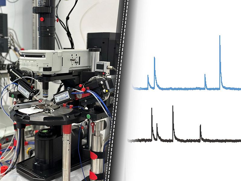
Patch-clamp electrophysiology is a powerful technique widely used in neuroscience research to investigate the electrical properties and activity of neural networks at the cellular level. It provides us with precise control over the measurement and manipulation of individual neurons, allowing for detailed characterization of their electrical behavior. Additionally, patch-clamp electrophysiology allows for the examination of synaptic communication between neurons, providing insights into the functional connectivity and network dynamics within neural circuits.
Deep Learning approach for EM
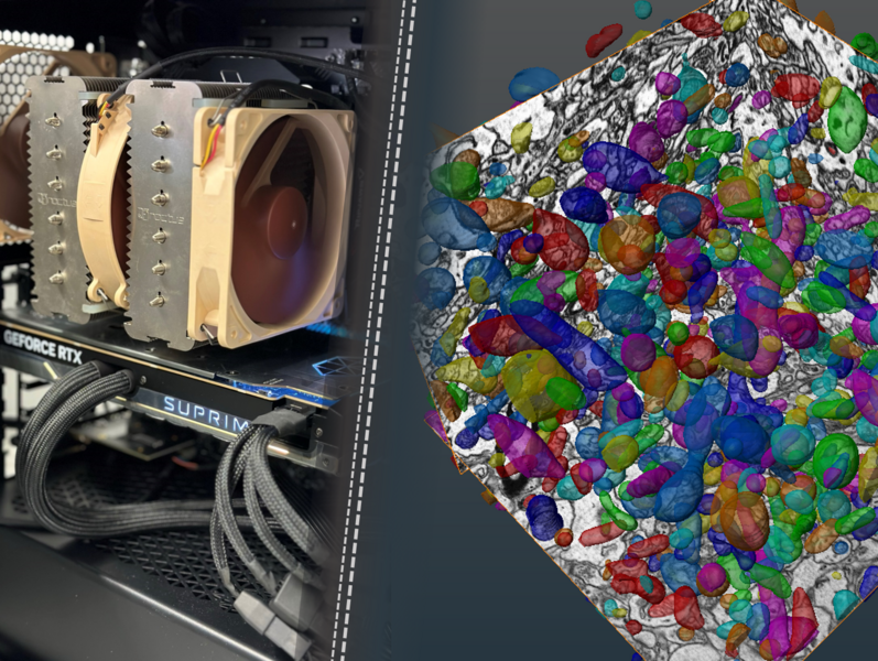
By leveraging machine learning algorithms, we can train models to recognize and categorize different components of the brain's ultrastructure. This involves feeding the model a vast amount of labeled data, such as electron microscopy images, where we have manually annotated the desired elements of interest, such as synaptic vesicles, postsynaptic densities or mitochondria. Once the model is trained, it can automatically analyze new, unlabeled images and accurately identify and label the ultrastructural elements present.
3D Neuron Reconstructions - Neurolucida
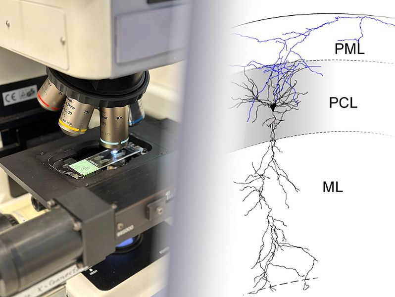
Overall, 3D Neurolucida reconstructions provide a powerful tool for investigating the complex morphology of individual neurons and understanding how their structure relates to their function. The detailed reconstructions contribute to our understanding of neuronal development, connectivity, and plasticity, ultimately advancing our knowledge of brain function and its implications in health and disease.
Focused Ion Beam Scanning Electron Microscopy (FIB-SEM)
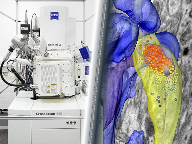
Focused Ion Beam Scanning Electron Microscopy (FIB-SEM) enables large volume 3D exploration of brain ultrastructure. Precise ion beam milling reveals layers while scanning electron microscopy captures images at nanoscale resolution. A resulting stack of high-resolution electron micrographs can be computationally reconstructed into a seamless 3D representation of various brain tissues including synapses, cytoskeleton and organelles. This technique is utilizing resources from the Core Facility Elektronenmikroskopie (CFEM).