Save the date: Night of the Arts on April 18, 2026
After more than 20 successful years of NIGHT OF THE MUSEUM, Düsseldorf's first NIGHT OF THE ARTS will take place on April 18, 2026.
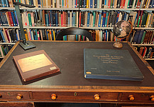
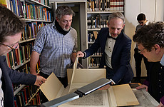
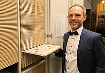
At the Cécile & Oskar Vogt Archive (Himmelgeister Straße 103, 40225 Düsseldorf), visitors can look forward to a unique insight into the world of brain research. The exhibition includes brain sections, images, graphics, and drawings, providing insight into brain research in the 20th and 21st centuries. The program includes a guided tour by a guest scientist and lectures on the use of historical material in modern brain research. The university's “Man and Death” graphic collection will also be presented. For those who want to learn about the brain and how it works, a workshop on its anatomy will be offered.
Further information can be found on the website.
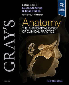
“Gray’s Anatomy” with a chapter by Katrin Amunts & Marco Catani
A special mark of distinction: As the successor to the late Prof. Karl Zilles, Prof. Katrin Amunts, together with Prof. Marco Catani, is responsible for the chapter on the "Cerebral hemispheres" in "Gray’s Anatomy". The 43rd edition of this standard reference work on human anatomy, first published in 1858, has just been released.
The chapter *Cerebral Hemispheres* by Amunts and Catani illustrates the significant advances in neuroimaging techniques: it includes, among other things, images from the Human Brain Atlas on the digital European research platform EBRAINS, as well as representations using 3D Polarized Light Imaging (3D‑PLI), which makes the trajectories of nerve fiber pathways in the brain visible.
"Science online" with Christian Schiffer
In the next episode of the Jülich series “Science Online”, Dr. Christian Schiffer will explain on Thursday, February 19, 2026, at 3 p.m., how researchers use artificial intelligence to decode the brain.
The INM-1 develops detailed anatomical models of the brain as well as three-dimensional maps that help researchers better understand its spatial organization. Artificial intelligence (AI) is used to analyze tens of thousands of high-resolution microscopic images of brain sections that have been digitized at INM-1 over many years. These images provide fascinating insights into the complex structure of nerve cells and their connections. In his lecture, Christian Schiffer will discuss the scientific, methodological, and technical challenges of this project and present current and future developments in AI methods for studying the structural organization of the brain.
The contemporary art world is also strongly influenced by this topic, as shown by the current exhibition of artist Shu Lea Cheang at the Ludwig Forum in Aachen. Since the 1990s, the artist has explored new digital technologies both as a subject and as a tool in various forms, including multimedia installations. Before Christian Schiffer’s lecture, curator Holger Otten will offer insights into the exhibition titled KI$$ KI$$.

- Copyright: Ludwig Forum
Obituary - Prof. Rita Süssmuth
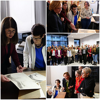
With deep sorrow, we bid farewell to Prof. Rita Süssmuth, former President of the German Bundestag, whose lifelong dedication to science, education, and society has left a lasting mark.
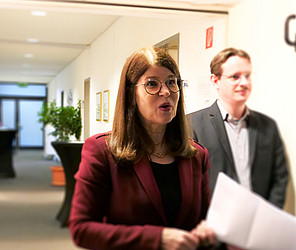
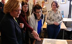
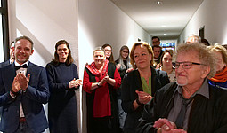
In January 2023, we had the honor of welcoming her as an honorary guest at the opening of the new Vogt Archive on Himmelgeisterstraße. Her visit was not only a special honor, but also a sign of appreciation for the archive and its importance to scholarship.
In the months following her visit, the collection of Cécile and Oskar Vogt was gradually transferred to its new location, digitized, and thus made it accessible to a broader community of researchers.
This digitization forms the foundation for integrating the Vogts’ historical brain mappings into the modern brain atlases of the 21st century - thereby carrying forward the legacy of an entire century of neurological research.
Rita Süssmuth’s commitment to science, education, and social participation will not be forgotten. Her visit, and her words of recognition and encouragement, were a source of motivation for us and continue to accompany our work at the archive.
Brain Awareness Week 2026
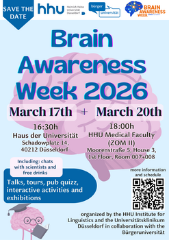
In March 2026, the Brain Awareness Week campaign will take place all around the world. The aim of the annual event is to arouse interest and enthusiasm for brain research. The global campaign is organized by the Dana Foundation and Federation of European Neuroscience Societies (FENS).
The C. and O. Vogt Institute will be presenting the latest research findings in brain research in an exhibition on March 17, 2026, starting at 4:30 p.m. at the Haus der Universität (Schadowplatz 14, 40225 Düsseldorf).
Participation is free of charge; registration is not required. Please find here further information.
New in Brain: The Vogt Collection in Düsseldorf
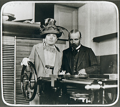
The year 2025 marks the 150th anniversary of Cécile Vogt's birth and the 155th anniversary of Oskar Vogt's birth. To mark this occasion, our institute’s director Katrin Amunts highlights the Vogts' scientific collection and their estate in the Vogt Archive for the renowned journal Brain. The conclusion: both represent a valuable resource for research in a broad spectrum ranging from the basic sciences and medicine to historical and ethical issues. The unique collection is now being fully cataloged and made available to the scientific community—as a source and source of inspiration for future research projects.
Katrin Amunts, The Vogt Collection: reactivating a treasure facilitating brain research, neurology and psychiatry, Brain, 2025; awaf365, https://doi.org/10.1093/brain/awaf365
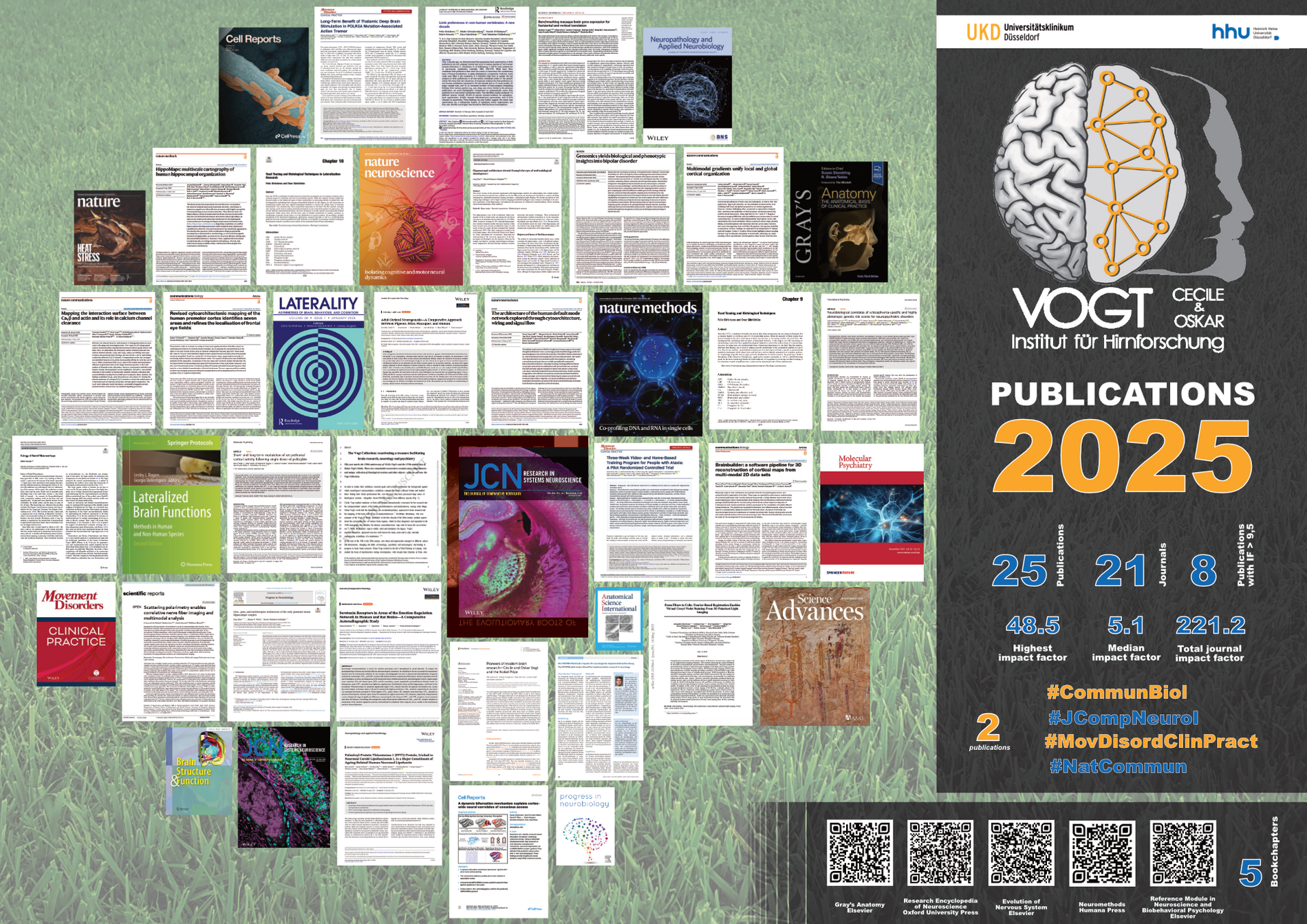
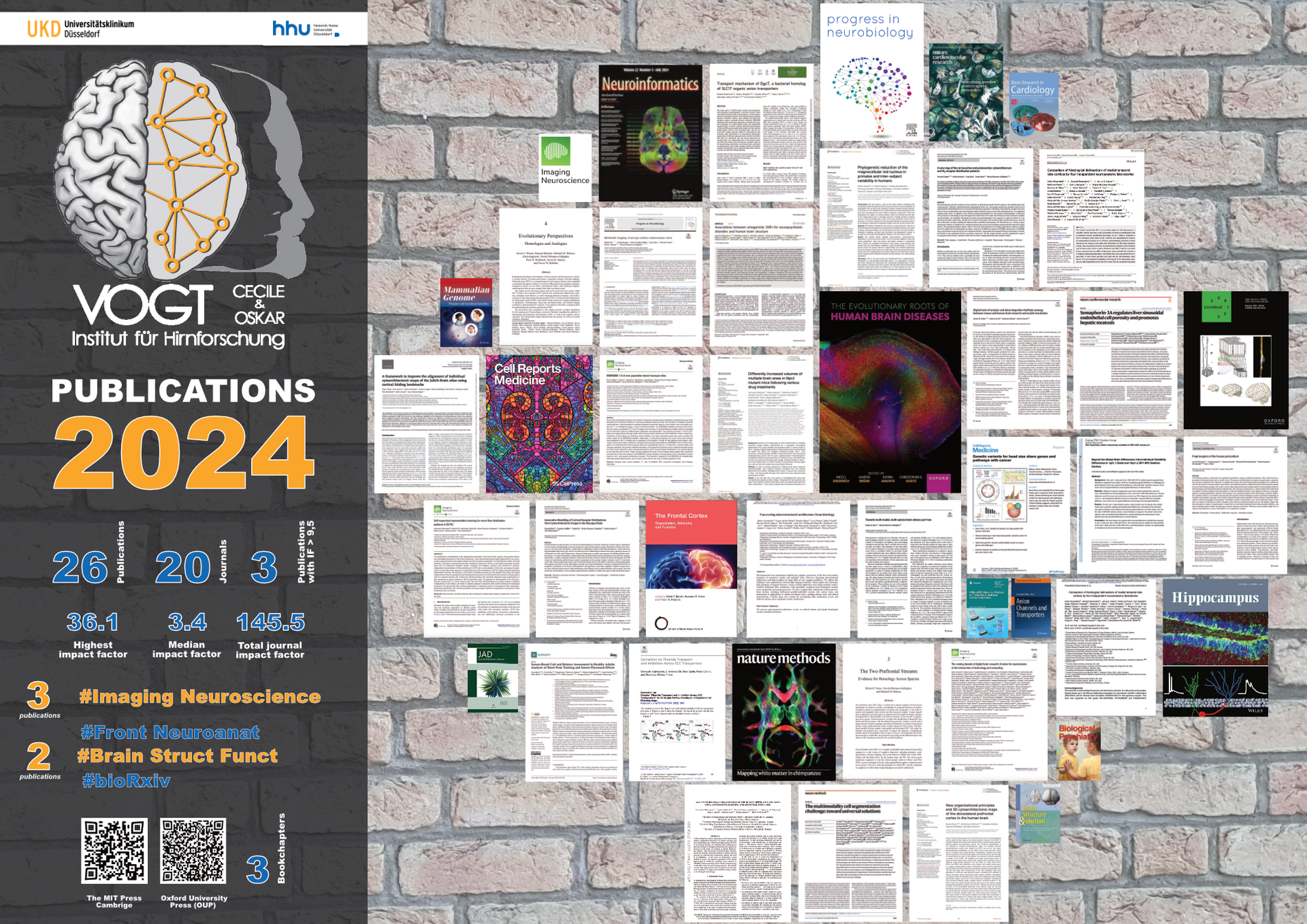
In 2025, our scientists have once again achieved amazing things, sharing their exciting results in leading neuroscience journals. A huge thank you to everyone who made these outstanding projects, collaborations, and publications possible!
We wish you all a joyful and peaceful holiday season, a relaxing break, and a fantastic start to the new year. May 2026 bring just as much success, inspiration, and groundbreaking discoveries!
From brain atlas to personalized model
Prof. Katrin Amunts was the head of the EU Flagship Human Brain Project. The project laid the foundation for a new brain atlas and personalized brain models, which are now available via EBRAINS. Such brain models can be used to plan tailored medical treatments. We spoke to her in this interview. You can find this interview here: https://www.fz-juelich.de/en/news/effzett/2025/from-brain-atlas-to-personalized-model
Premotor Cortex Remapped: Seven Subareas and Functional Distinction
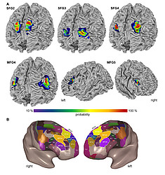
Researchers from our institute and the Institute of Neuroscience and Medicine (INM-1) have remapped the human premotor cortex, identifying seven clearly distinguishable subareas. The new histologically high-resolution maps show how the different regions are anatomically delineated. This new subdivision helps clarify the functional differences between these regions. The new maps are available in the Julich Brain Atlas, a core component of EBRAINS—the European digital research platform for neuroscience. The study has now been published in Communications Biology.
Please find here further information.
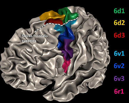
Copyright: Ruland, S. et al. Commun Biol 8, 1143 (2025). https://doi.org/10.1038/s42003-025-08528-4
NRW Academy: Brain Research, Media Art, and Literature in Dialogue
“Between Image and Language – Thinking in the Telematic Society” – under this title, the NRW Academy of Sciences and Arts in Düsseldorf invited guests on Tuesday, November 25, 2025, to an evening of lectures and discussion. After an opening talk by Professor Katrin Amunts, an extended conversation followed, featuring the neuroscientist herself along with the writer Marion Poschmann and media theorist Professor Siegfried Zielinski. The evening was moderated by Mischa Kuball, artist and professor of media art.
Please find further information here.
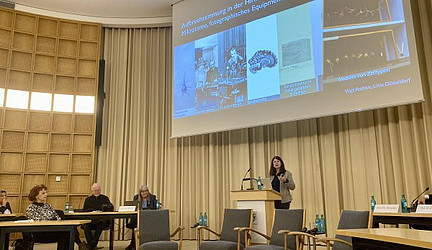
Copyright: Forschungszentrum Jülich / Peter Zekert
The Julich Brain Atlas and the “Telematic Society”
“Between Image and Language – Thinking in the Telematic Society” is the title of the lecture by Professor Katrin Amunts on Tuesday, November 25, 2025, at the North Rhine-Westphalian Academy of Sciences, Humanities and the Arts in Düsseldorf. The background is the utopia of a “telematic society,” conceived by media philosopher Vilém Flusser more than forty years ago. In such a society, human and technical communication systems are inseparably intertwined. According to this utopian vision, a world so thoroughly digitalized would itself digitalize human thought and radically transform the symbols of human exchange.
The event begins at 6:30 p.m. at the Academy’s headquarters, Palmenstraße 16, 40217 Düsseldorf. Registration: anmeldung@awk.nrw.de .
Please find further information on their website.
Why two pioneers of brain research never received the Nobel Prize
A new article in Frontiers in Neuroanatomy examines the scientific legacy of Cécile and Oskar Vogt. Their joint work shaped modern brain research—yet they never received the Nobel Prize, despite numerous nominations. Authors Nils Hansson, Heiner Fangerau, Fabio De Sio, Ursula Grell, and Katrin Amunts draw on archival sources from the Nobel Forum in Sweden and the Vogt Archive in Düsseldorf to understand why the research couple was nominated repeatedly over decades, yet the Nobel Prize Committee always decided otherwise. The article also reflects on how the Vogts' work lives on in modern neuroscience. The article was written in collaboration with researchers from the C. u. O. Vogt Institute for Brain Research, the Vogt Archive, and the Institute for the History, Theory, and Ethics of Medicine at Düsseldorf University Hospital.
https://www.frontiersin.org/journals/neuroanatomy/articles/10.3389/fnana.2025.1679993/full
Nils Hansson, Heiner Fangerau, Fabio De Sio, Ursula Grell and Katrin Amunts (2025). Pioneers of modern brain research—Cécile and Oskar Vogt and the Nobel Prize. Front. Neuroanat. 19:1679993. doi: https://doi.org/10.3389/fnana.2025.1679993
Poster Award for Denise Kalbe
Denise Kalbe received third prize for her poster at the “BMFZ Meeting – 6th Symposium on Neurodegenerative Diseases” held in mid-November at the Haus der Universität in Düsseldorf. Under the title “Three-Dimensional Polarised Light Imaging in Neurodegeneration: Focus on MSA-C,” the poster demonstrated how 3D-Polarized Light Imaging (3D-PLI) can be used to study neurodegenerative diseases. The technique visualizes the orientation and integrity of nerve fibers in brain sections. By exploiting the birefringence of brain tissue, it provides high-resolution insights into the myelin sheaths of nerve fibers and reveals their damage.
Further information can be found here.
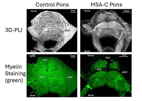
Copyright: Denise Kalbe
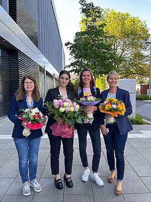
Congratulations to Zien Zhang!
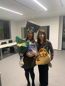
We are delighted that Zien Zhang has successfully completed her doctoral thesis entitled “Cytoarchitectonic mapping of five new areas in the anterior lateral prefrontal cortex” at the Medical Faculty of Heinrich Heine University Düsseldorf. In her thesis, five new cytoarchitectonic areas in the lateral prefrontal cortex were identified, the maps are available at the Julich-Brain Atlas —an exciting contribution to a better understanding of the structure and function of the human brain.
The entire institute congratulates her warmly and wishes her all the best for her future scientific career!
New Publication Alert
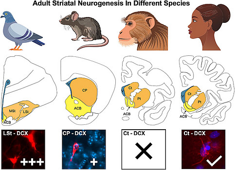
Can the adult brain still generate new neurons, even beyond the hippocampus? A new study from our colleagues takes a closer look at this fascinating question across species - from pigeons and mice to macaques and humans.
In their paper, “Adult Striatal Neurogenesis - A Comparative Approach Between Pigeons, Mice, Macaques, and Humans” the team quantitatively analyzed neurogenic markers in different striatal regions. Their findings reveal remarkable differences: pigeons show a much higher degree of neuronal plasticity and stem cell activity compared to mammals. Interestingly, signs of persistent neuronal plasticity were also observed in the human caudate nucleus, offering new perspectives on how the adult human brain might maintain its ability to adapt and renew itself.
These insights highlight birds as promising models for studying adult neurogenesis and could ultimately help us better understand the mechanisms of neuronal plasticity in the human brain.
Read the full study here: https://onlinelibrary.wiley.com/doi/10.1002/cne.70107
Further Award for Nicola Palomero-Gallagher
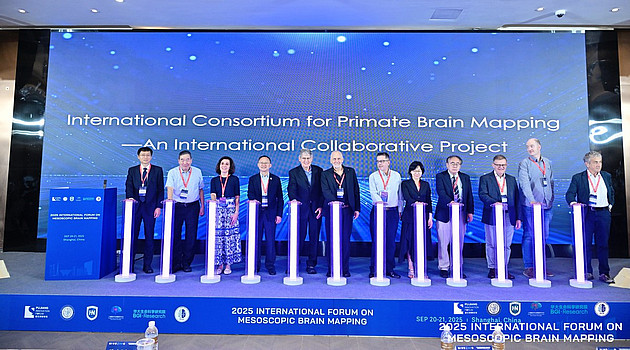
At this year's "International Forum on Mesoscopic Brain Mapping" held in September in Shanghai, the "International Consortium for Primate Brain Mapping" was founded. The members elected Associate Professor Nicola Palomero-Gallagher (INM-1) along with Professor Alan Evans (McGill University) as deputy chairs of the scientific advisory board.
The main objective of the consortium is to understand, over the next ten years, the fundamental principles by which the structure of primate brains enables cognitive and perceptual functions. To achieve this, an international network of researchers will be established to work on the first mesoscopic atlas of primate brains.
Copyright:
— 2025 International Forum on Mesoscopic Brain Mapping
"HippoMaps" simplifies hippocampus research
Despite the important role of the hippocampus in many cognitive processes of the human brain, there has so far been no unified system to record, jointly present, and analyze the structure and function of this region using different measurement methods. For this reason, an international team of scientists has developed "HippoMaps" within the framework of the German-Canadian HIBALL project (Helmholtz International BigBrain Analytics and Learning Laboratory): a freely accessible software toolbox including an online database, specifically for deep learning-based integration of image data from various sources and resolutions, as well as for the analysis and mapping of the hippocampus. The aim is to better understand the relationships between its structure and function in both healthy and diseased brains. In a recent study published in Nature Methods, they present "HippoMaps".
Further information can be found here.
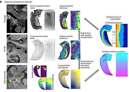
— DeKraker, J. et al. HippoMaps: multiscale cartography of human hippocampal organization. https://doi.org/10.1038/s41592-025-02783-3
Max Planck School of Cognition
Application Portal now open!
The current application cycle for the doctoral program started again on September 1st 2025. The application portal will remain open from this day until December 1st 2025 for intake 2026! The program starts each year on September 1st.
Please submit your application at the application portal.
Hector Research Career Development Award
New application phase from 01.09.-30.10.2025
The new application phase for the Hector Research Career Development Award (Hector RCD Award) opened on September 1. Qualified candidates who are in the transition phase between postdoc and professorship can apply again until October 30, 2025.
The award is specifically aimed at W1 professors and junior research group leaders and is endowed with €25,000 in funding.
Please find here further information.
Hippocampal Research: History, Methods, and Perspectives for the Neurosciences
A recent review by researchers at the Institute of Neurosciences and Medicine (INM-1) examines the development of methodology—and thus the progress—in the neurosciences using the hippocampus as an example, the brain region responsible for learning, memory, and spatial orientation. For more than 150 years, innovative methods coupled with meticulous neuroanatomical analyses have enabled increasingly detailed insights into the structure and function of the brain. The study, now published in Anatomical Science International, concludes that future methodological advances in brain research must necessarily be comparative and interdisciplinary—incorporating the expertise of physicists, computational neuroscientists, and, above all, classical neuroanatomists.
The work of the Jülich scientists focuses on the structural organization of the hippocampus in both the human brain and in animal models frequently used in neuroscience research. It addresses various components such as cellular structure, molecular diversity, and connectivity. A broad overview is provided of the different research methods. It begins with the Golgi staining technique, developed in the 1870s, and covers further methods such as immunohistochemical staining and receptor autoradiography. Modern invasive and non-invasive tractography techniques, such as magnetic resonance imaging (MRI), round off the overview.
The authors also look to the future with the digitization of the neurosciences. To analyse the massive, highly complex data sets emerging from research, neuroscientists increasingly rely on artificial intelligence (AI) methods. For instance, deep learning algorithms and specially prepared training data from neuroanatomy have been used to automatically segment the entire "BigBrain"—an extremely high-resolution 3D model of a human brain—into its individual layers.
New AI-supported approaches are also being applied in hippocampal research. Here, state-of-the-art imaging data are combined with statistical learning techniques to capture the fine neural circuitry within the hippocampus. The results align with classical anatomical subdivisions.
The researchers attribute particular significance to the comparative approach: the hippocampus has evolved over time in terms of size, neuronal connectivity, and synaptic plasticity. Comparing different species makes it possible to better understand structural adaptations and to improve the transfer of findings from animal experiments to humans—an important step in developing targeted therapies for neurological and psychiatric diseases.
To analyse the resulting large and complex data sets, the researchers propose the "Common Space" concept. Originally developed to overcome methodological limitations between different species, this method allows integration of data from various measurement techniques—from molecular profiles and connectivity analyses to functional imaging—within a unified analysis framework. Applied to the hippocampus, this approach could help integrate high-resolution anatomical data, temporally resolved data, and computational models, thus enabling comprehensive deciphering of the relationship between its microstructural organization and large-scale brain dynamics.
Original publication:
Zhao, L., Palomero-Gallagher, N. Hippocampal architecture viewed through the eyes of methodological development. Anat Sci Int (2025). https://doi.org/10.1007/s12565-025-00878-7
The press release can also be found here.
Contact: apl.Prof. Dr.rer.nat. Nicola Palomero-Gallagher, Phone: +49 2461/61-4790, E-Mail; Press contact: Erhard Zeiss, Phone: +49 2461/61-1841, E-Mail
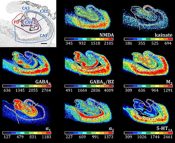
With new brain maps, the eye fields can be located more precisely

Copyright:
Ruland, S.H., Sigl, B., Stangier, J. et al., Commun Biol 8, 1143 (2025). https://doi.org/10.1038/s42003-025-08528-4
Researchers have conducted a detailed study and re-mapping of the so-called premotor cortex in the brain, which controls movements and cognitive processes. The new maps have enabled the precise localization of the anatomical correlates of the so-called eye fields for the first time. These new findings enhance imaging precision and may help improve clinical applications. The study by researchers from INM-1 and the Cécile and Oskar Institute for Brain Research at the University Hospital Düsseldorf has now been published in Communications Biology.
Further information can be found here.
Deep brain stimulation proves effective in rare genetic motor disorder

Copyright:
— Minnerop, M. et al.
In the journal Movement Disorders Clinical Practice, scientists of the Clinical Neuroanatomy research group at Jülich's INM-1, in cooperation with the Centers for Movement Disorders and Neuromodulation as well as Functional Neurosurgery and Stereotaxy at Düsseldorf University Hospital, have presented a new case report on the effect of deep brain stimulation in a patient with a rare genetic motor disorder. Caused by the mutation of a gene called POLR3A, this disease often results in a pronounced so-called action tremor, which disrupts any voluntary movement of the affected person through severe tremor.
Further information can be found here.
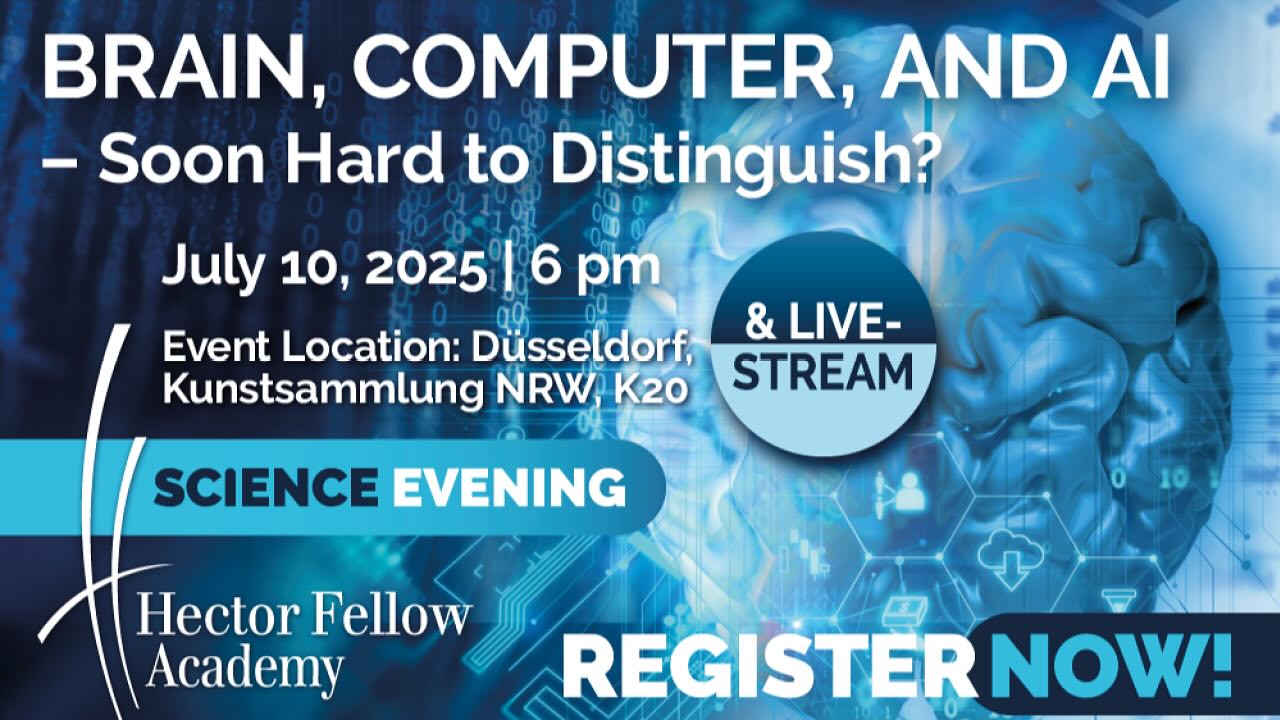
Successful science evening of the Hector Fellow Academy
“Brain, computer and AI - soon hard to distinguish?” - Prof. Katrin Amunts (Cécile and Oskar Vogt Institute for Brain Research/Forschungszentrum Jülich), Prof. Rainer Goebel (Maastricht University) and Prof. Thomas Lippert (Forschungszentrum Jülich/Goethe University Frankfurt) provided answers to this question at the Hector Fellow Academy Science Evening in Düsseldorf on Thursday, July 10, 2025. In their talks, Amunts, scientific organizer of the evening, and her two colleagues presented to the audience in the lecture hall of the Kunstsammlung NRW and via livestream how supercomputers generate artificial intelligence, what we still want to learn from the structure of the brain and how the question can be answered from the perspective of cognitive neuroscience.
Further information can be found here.
A recording of the science evening is available on the Hector Fellow Academy YouTube channel: https://youtu.be/auBNu_u8P8M

Copyright:
— Hector Fellow Academy
EBRAINS Summit 2025 from 8-11 December 2025 in Brussels
Join Europe’s leading minds in neuroscience, innovation, and policy for the EBRAINS Summit 2025 – Transforming Brain Research and Medicine – live in Brussels on 8-11 December. Please find here the registration link: https://summit2025.ebrains.eu/register

Hector Fellow Symposium at K20 Düsseldorf
Join the Hector Fellow Academy Science Evening on July 10 in Düsseldorf! Registration is open. Register here.
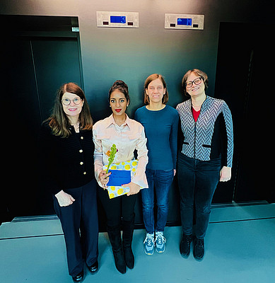
Congratulations, Dr. Manuel Dietermann!
We are proud to congratulate our doctoral candidate Manuel Dietermann on the successful completion of his PhD!
Under the academic supervision of Prof. Dr. Dr. h.c. Katrin Amunts and Prof. Dr. Dr. Svenja Caspers, Manuel has made a significant contribution to brain research through his excellent scientific work.
The entire team at the Cécile & Oskar Vogt Institute of Brain Research is celebrating this important milestone with you.
We are proud of you!
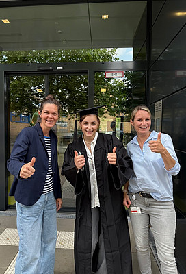
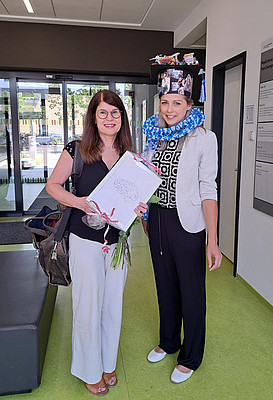
She did it - now Miss Dr. Meckenstock!
Yesterday, Laura Meckenstock successfully defended her PhD with a fascinating topic exploring the influence of different learning conditions on adult neurogenesis in the hippocampus of pigeons.
We are incredibly proud of you, especially your supervisors PD Dr. Christina Herold and PD Dr. Julia Mehlhorn.
Now it’s time to breathe a sigh of relief, celebrate, and enjoy the moment!
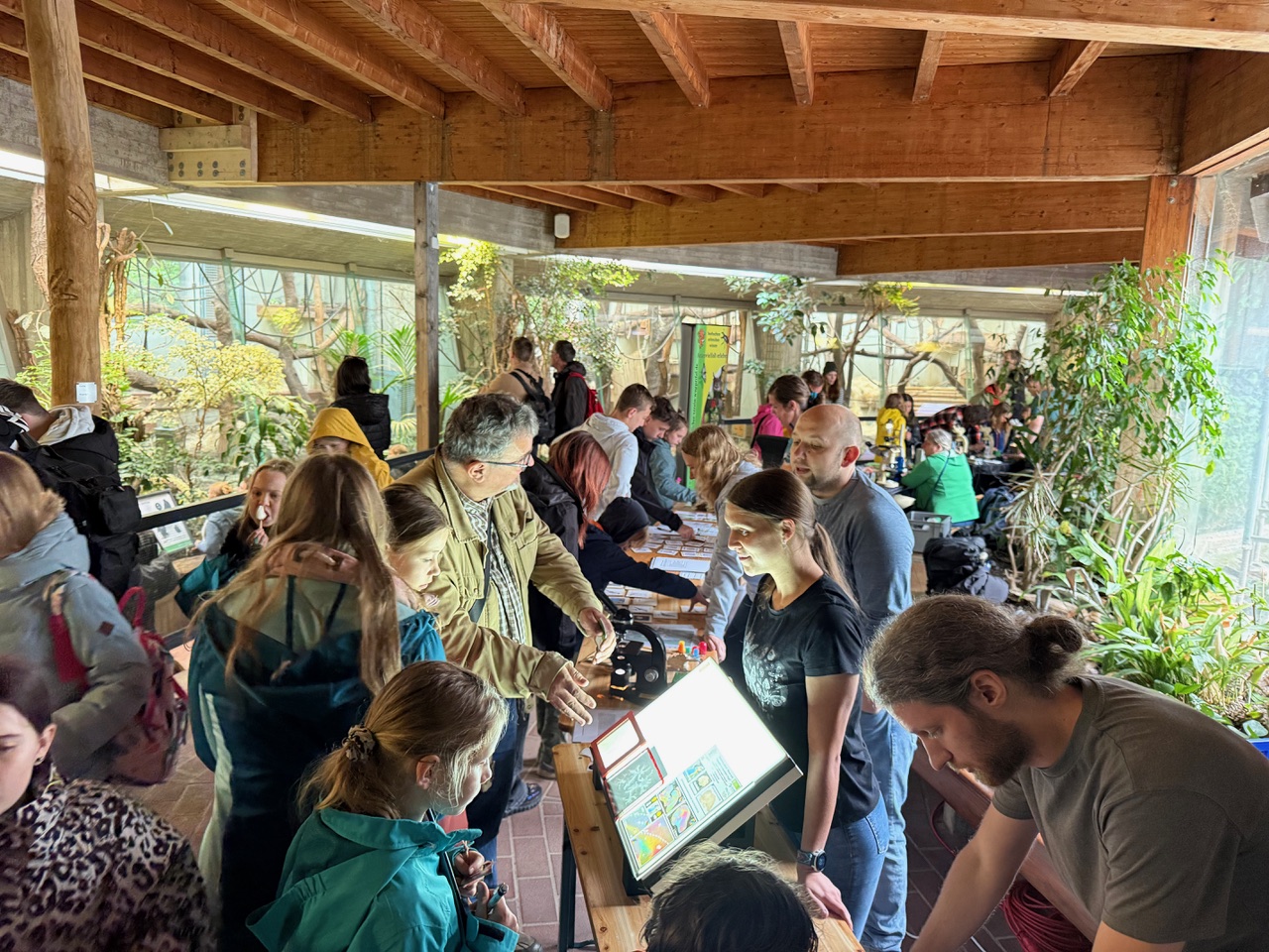
Zoo Day at the „Grüner Zoo Wuppertal" – May 24, 2025
For the third year in a row, we were excited to take part in this special day as returning guests. Each year, the „Grüner Zoo Wuppertal" invites visitors to explore the world of nature conservation, species protection, and climate awareness through a wide range of engaging exhibits. We are always happy to participate - bringing the colorful world of neuroscience to life in a funny and interactive way for visitors of all ages. Together with Prof. Dr. Markus Axer (University of Wuppertal and Research Center Jülich), we supported our long-standing collaboration partner by offering hands-on activities. As in previous years, guests were invited not just to observe, but also to touch, play, puzzle, guess, use microscopes, and experiment.
"Vimoto," the silverback gorilla, observed us with his usual calm as we engaged with curious zoo visitors about the brains of his species - comparing them to those of orangutans, guenons, kangaroos, domestic pigs, guinea pigs, rats, birds, frogs, crocodiles, and of course, humans. We're especially thankful to the founders of our institute, whose comprehensive collections allow us to explore and share insights into the brains of such a wide range of species. There were plenty of enjoyable conversations and surprising discoveries throughout the day. Some guests discovered hidden talents at the polarimeter or showed remarkable memory skills during the game. We hope we were able to pass on some of our enthusiasm for the topic to many young and adult visitors to our exhibition booth.
Our heartfelt thanks go once again to the entire team at the "Grüner Zoo Wuppertal" for the outstanding organization and smooth execution of the event - especially to our cooperation partner Dr. Dominik Fischer. We're already looking forward to next year!
Author: PD Dr. Christina Herold
It is well known that around 90% of humans are right-handed, while approximately 10% prefer using their left hand. But what about animals?
We're excited to share a brand-new scientific paper by our colleague Felix Ströckens and co-authors: Limb preferences in non-human vertebrates: A new decade in the journal of Laterality.
More than ten years after their landmark 2013 study, the team of researchers from our C. & O. Vogt Institute of Brain Research at HHU Düsseldorf, the Ruhr University Bochum, and MSH Medical School Hamburg, reviewed the scientific literature on limb usage in non-human vertebrates. They found that many vertebrate species also exhibit limb-use asymmetries - just like humans do. Their updated analysis spans 172 species across all non-extinct vertebrate orders, revealing that limb asymmetries are far more widespread than previously thought.
- 39.53% of species show population-level limb asymmetries
- 32.56% show individual-level asymmetries
- only 27.91% show no asymmetry at all
These findings clearly indicate that handedness is not unique to humans. Instead, it appears across a wide range of animal species and may have evolved in conjunction with the development of bilateral limbs.
Ströckens, F., Schwalvenberg, M., El Basbasse, Y., Amunts, K., Güntürkün, O., & Ocklenburg, S. (2025). Limb preferences in non-human vertebrates: A new decade. Laterality, 1–46. https://doi.org/10.1080/1357650X.2025.2499049
Master’s program Translational Neuroscience at HHU Düsseldorf
The application process for our master's program Translational Neuroscience will start on 15th May 2025. The deadline will end on 15th July 2025.
Further information can be found here: https://www.translationalneuroscience.hhu.de/
We are looking forward to your application!
Congratulations on your PhD, Dr. Karsli!
Yesterday, our doctoral candidate Erhan Karsli successfully defended his dissertation at the Medical Faculty of Heinrich Heine University Düsseldorf. The entire team at the C.&O. Vogt Institute of Brain Research congratulates you with great pride and joy on this significant milestone.
We wish you much success, exciting challenges, and above all, joy in everything you do as you move forward. All the best for your future, dear Erhan!
From Poster Presenter to Symposium Organizer: Our Journey at Göttingen Meeting 2025
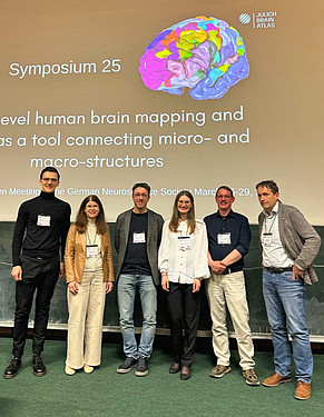
Visiting a scientific conference as a PhD student – presenting a poster, joining discussions, and learning from leading researchers - feels already like an achievement. But organizing a full symposium as early-career researchers? That was something we never imagined ourselves doing. Over a year ago, encouraged by our colleague Felix Ströckens, and by the high trust and support of our supervisor Prof. Katrin Amunts, we accept that challenge. Together, we start to organize a symposium as part of the 16th Göttingen Meeting of the German Neuroscience Society (NWG), which took place from March 26-29, 2025.
Our session, “Multilevel Human Brain Mapping and Atlas as a Tool Connecting Micro and Macrostructures”, brought together 70–80 participants. We were honoured to feature inspiring keynote talks: Prof. Katrin Amunts discussed the evolution of brain mapping and the role of the Julich Brain Atlas in studying structure-function relationships. Prof. Timo Dickscheid introduced the Siibra tool suite and its capabilities for integrating multimodal brain data, and Prof. Tim Salditt shared advances in 3D virtual histology using synchrotron-based X-ray tomography. We were also proud to present our own research: Alexey shared findings on high-resolution 3D mapping of the human hypothalamus and its subdivisions, and Nataliia followed with her presentation on the cytoarchitectonic delineation of Broca’s region, highlighting new subdivisions in areas 44 and 45. Carla Hogrebe, our fellow PhD student, was also at the Göttingen Meeting 2025. She presented her cytoarchitectonic mapping of the human temporal pole during the poster sessions just the day before our symposium. She and Felix were there to support us – having familiar faces in the audience was comforting.
One of our favourite moments? That special moment when active discussion continued long after the official session ended, with participants approaching speakers to exchange ideas and explore collaboration opportunities. We received a lot of positive feedback and even one suggestion to join the annual science festival Pint of Science – which we had seriously considered. The warm spring weather in Göttingen made the conference enjoyable, with evening walks through the historic city providing a relaxing break after busy days of scientific exchange and networking. Organizing a symposium was challenging, yes – but deeply rewarding. We’re incredibly grateful for the support of our colleagues at the Cécile & Oskar Vogt Institute of Brain Research Düsseldorf and INM-1 at Research Centre Jülich, and for everyone who helped bring this to life.
We left Göttingen with a sense of satisfaction and inspiration, grateful for the opportunity to contribute to and learn from such a vibrant scientific community. To fellow PhD students considering taking a similar step: DO IT! You’re more ready than you think.
Nataliia & Alexey
Authors: Nataliia Fedorchenko - PhD fellow of the Max Planck School of Cognition and Alexey Chervonnyy, PhD fellow of the Hector Fellow Academy
New: WDR Podcast about brain researcher Oskar Vogt
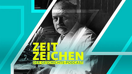
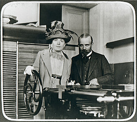
A new episode in the podcast series WDR Zeitzeichen titled “Oskar Vogt: The Measurement of the Brain” was released on April 6, 2025, marking the 155th birthday of the pioneering brain researcher. Author Daniela Wakonigg traces the life and achievements of Oskar Vogt and his wife Cécile, who worked closely together for decaerdes in groundbreaking neuroscience research. Prof. Katrin Amunts, Director of the Cécile and Oskar Vogt Institute of Brain Research at University Hospital Düsseldorf and of the Institute Structural and Functional Organisation of the Brain (INM-1), and Dr. Manuel Marx, Scientific Coordinator of the Vogt Archive, are among the featured speakers. The 15-minute episode explores how the Vogts became pioneers of localization theory in brain research and what insights they gained from their examination of Lenin's brain. It also delves into their experience during the Nazi era, their continued work at a private research institute in Neustadt-Titisee, and the recent relocation and digitization of their archive by the Düsseldorf Brain Research Institute. Copyright image: Picture Alliance/Ullstein-Bild, Author: Erhard Zeiss, translated by: Manuel Marx
https://www1.wdr.de/radio/wdr5/sendungen/zeitzeichen/zeitzeichen-oskar-vogt-100.html
New exhibition "Brains" at Senckenberg Museum in Frankfurt
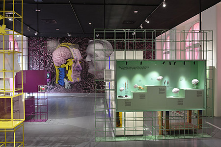
An interview on current brain research and the Jülich Brain Atlas with Prof. Katrin Amunts, Director of the Cécile and Oskar Vogt Institute for Brain Research at University Hospital and Heinrich Heine University Düsseldorf and of the Institute for Structural and Functional Organization of the Brain (INM-1), Research Centre Jülich, is part of the new permanent exhibition “Brains”, which has now been opened at the Senckenberg Research Institute and Natural History Museum in Frankfurt. The museum displays over 120 exhibits on the topic of human and animal brains – their diversity, evolutionary development and changes over the course of a lifetime – in an area of 200 square meters. The Cécile and Oskar Vogt Institute in Düsseldorf has provided nerve cell models, stained sections of the brain and spinal cord, and a drawing of cell layers.
Further information can be found here.
University Information Day on 14th June 2025 at HHU Düsseldorf
On Saturday, June 14, 2025, from 10:00 am to 3:00 pm, Heinrich Heine University Düsseldorf and Düsseldorf University of Applied Sciences will be presenting the range of courses available in Düsseldorf and providing support in choosing a course of study at the University Education Information Day.
We invite students, together with their parents and teachers, to come to the campus of Heinrich Heine University to find out about the wide range of degree programs and campus life in Düsseldorf.
Save the date: HFA Science Evening on July 10, 2025 in Düsseldorf
This year, Professor Amunts is hosting the Hector Fellow Academy's Science Evening under the title “Brain, Computer and AI: Soon hard to distinguish?” It will take place on July 10, 2025 at 6 pm at K20 in Düsseldorf.
EBRAINS tutorials and users day in Heidelberg on 12th March 2025
The EBRAINS tutorials and users day will take place in Heidelberg on 12th March 2025. The Workshop is aimed at two audiences: newcomers to EBRAINS who are interested in learning how to use the tools and services available on the research infrastructure, and seasoned EBRAINS users who would like to attend advanced tutorials and user discussion groups with developers.
Further information can be found on their website.
Night of Libraries Düsseldorf
On April 4, 2025, the University and State Library Düsseldorf is hosting the Night of Libraries. This year's motto: “Knowledge. Share. Discover”. Short lectures and hands-on activities will take place from 6 pm to 11 pm. Among others, our colleague PD Dr. Christina Herold will give a lecture on “A neuron wants to know - How knowledge is created in the brain!”. We look forward to your visit.
Further information about the event and the program can be found on the ULB website.

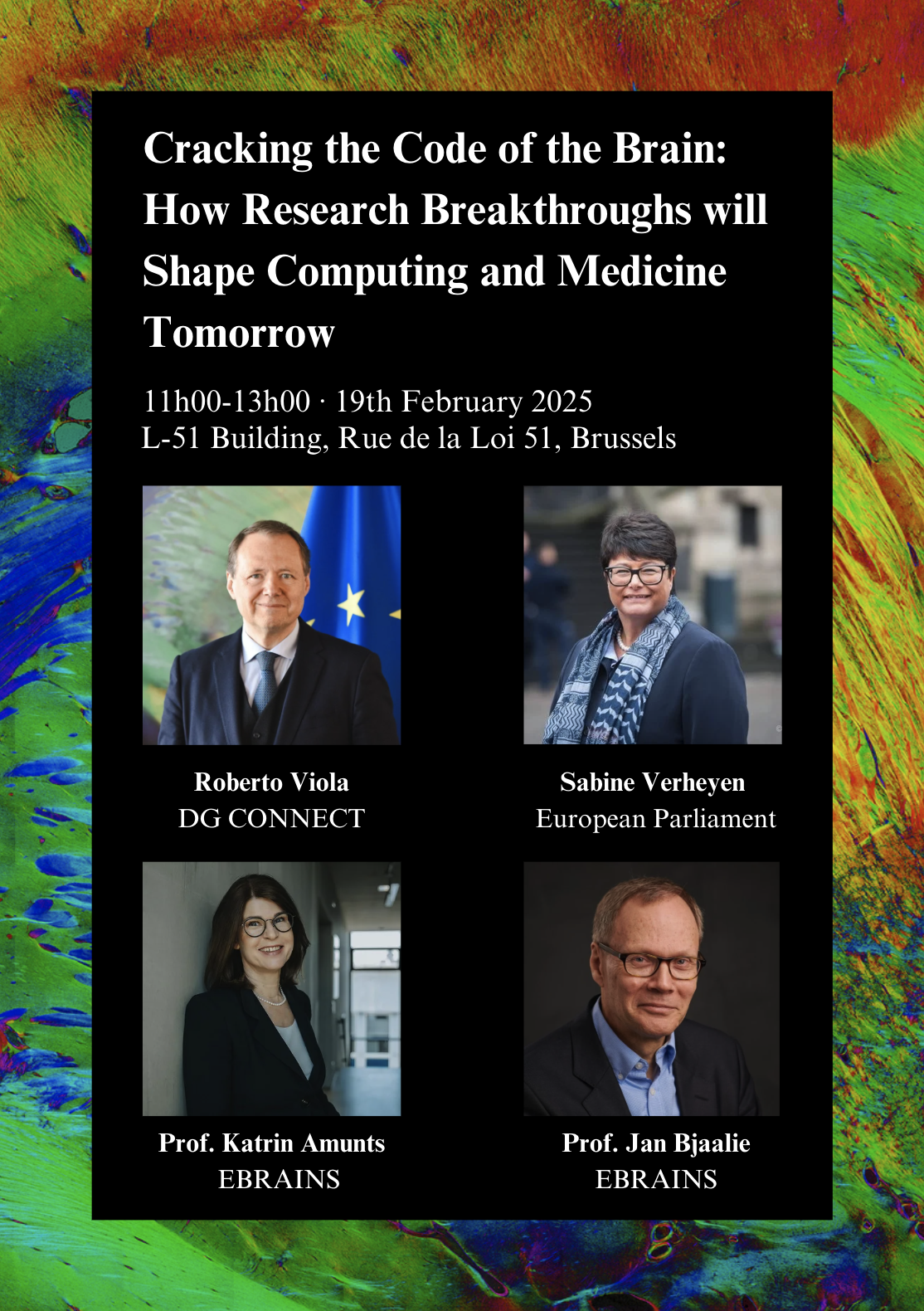
Next week in Brussels, the success of the Human Brain Project and the ongoing impact of the EBRAINS Research Infrastructure will be celebrated with the opening of the HBP-EBRAINS exhibition at the L-51 Building of the European Commission on February 19, 2025.
The event will be inaugurated by Roberto Viola, Director-General of DG CONNECT at the European Commission, and Sabine Verheyen, First Vice-President of the European Parliament. Following the opening remarks, EBRAINS Joint-CEO Katrin Amunts and EBRAINS Chief Infrastructure Officer Jan Bjaalie will deliver keynote presentations.
More details about the event: https://www.ebrains.eu/news-and-events/hbp-ebrains-exhibition-at-european-commission-to-open-in-february-2025

Save the date: 3rd Action Day at the Green Zoo Wuppertal on May 24, 2025.
This year, the C. and O. Vogt Institute of Brain Research is once again participating in the Action Day at the Green Zoo Wuppertal under the theme “Nature Conservation, Species Protection, and Climate Protection.” The event will take place on May 24, 2025, from 1 PM to 6 PM. For the third time, the Action Day is being organized in cooperation with the Wuppertal Institute. Professor Axer (INM-1, Research Center Jülich, and Bergische Universität Wuppertal) and PD Dr. Herold (C. and O. Vogt Institute of Brain Research, University Hospital, and HHU Düsseldorf) will present their work at an information booth. Exciting interactive activities are also planned. Come and join us!
Katrin Amunts receives honorary doctorate from Maastricht University
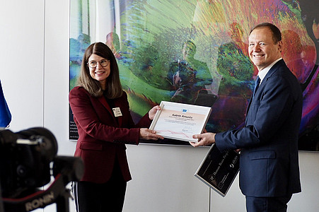
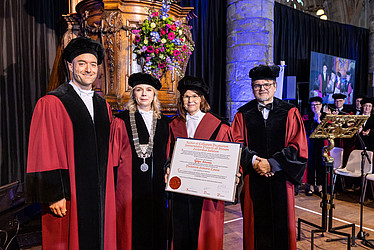
Maastricht/Jülich, February 3, 2025 - On Friday, January 31, 2025, Maastricht University awarded an honorary doctorate to Prof. Katrin Amunts in recognition of her outstanding and inspiring scientific achievements. The world-renowned neuroscientist received the certificate in a ceremony with Rector Prof. Pamela Habibović in the Sint-Janskerk. Every year, the university commemorates its foundation in 1976 with the ceremonial “Dies Natalis”.
Katrin Amunts is Director of the Institute of Neuroscience and Medicine (INM-1) at Forschungszentrum Jülich and Director of the C. and O. Vogt Institute for Brain Research at Heinrich Heine University Düsseldorf. From 2016 until its successful completion in 2023, the brain researcher was Scientific Director of the European flagship “Human Brain Project” (HBP). Katrin Amunts and her research team were able to map the human brain on an unprecedented scale using extremely high-resolution and data-intensive methods. This was achieved in an interdisciplinary collaboration with experts from the Jülich Supercomputing Center (JSC). The results of this research are combined in a globally unique 3D brain atlas, which is openly accessible to scientists from all over the world in EBRAINS, the digital platform for brain research developed at the HBP.
The laudators Prof. Rainer Goebel and Prof. Alard Roebroeck from the Faculty of Psychology and Neuroscience both emphasized the vision, pioneering spirit and perseverance with which Katrin Amunts made groundbreaking advances in neuroscience over the past decades. Alard Roebroeck referred to the multimodal, multiscale Julich Brain Atlas, which has benefited brain research worldwide. Rainer Goebel emphasized the scientist's close collaboration with Maastricht University, which led to the establishment of a new degree course. There, future brain researchers will be trained at the interface of psychology, neuroscience, medicine and artificial intelligence.
Katrin Amunts' work has received numerous awards. The scientist is a member of the German National Academy of Sciences Leopoldina, a full member of the German National Academy of Sciences and Engineering (acatech) and a full member of the North Rhine-Westphalian Academy of Sciences, Humanities and the Arts. From 2012 to 2020, she was one of 26 members of the German Ethics Council. Katrin Amunts was awarded the prestigious Hector Science Prize in 2022, the same year the then North Rhine-Westphalian Science Minister Isabel Pfeiffer-Poensgen presented her with the Cross of Merit 1st Class of the Order of Merit of the Federal Republic of Germany. In 2023, she received the international Justine and Yves Sergent Award.
Recording of the ceremony (awarding of the honorary doctorate from min. 47:00) https://www.youtube.com/watch?v=OCRLZw38aeE
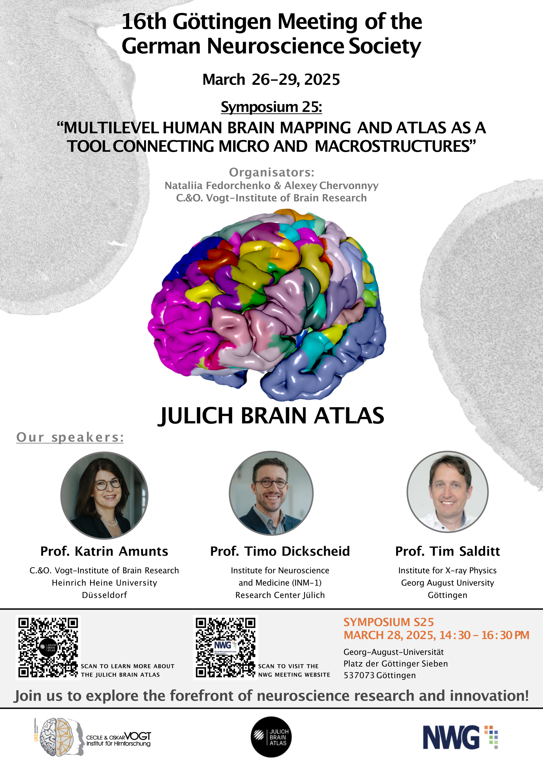
Symposium Announcement:
“MULTILEVEL HUMAN BRAIN MAPPING AND ATLAS AS A TOOL CONNECTING MICRO AND MACROSTRUCTURES”
The C. & O. Vogt Institute of Brain Research is pleased to announce its participation in the 16th Göttingen Meeting of the German Neuroscience Society. As part of this event, our colleagues will organize the symposium, “Julich Brain – A Multimodal 3D Atlas of Cortical and Subcortical Areas” on 28 March 2025, from 14:30 to 16:30, in Göttingen.
About the Symposium
Understanding cognitive networks requires precise knowledge of brain organization, including the areas and nuclei that form these networks. Many brain atlases focus on surface features, such as sulci and gyri, leaving subcortical structures – critical hubs in large-scale networks – underrepresented. Moreover, sulcal and gyral patterns often do not correspond to cortical area boundaries, which are only visible at the microscopic level. This challenge is compounded by the significant inter-individual variability of the human brain.
The Julich Brain Atlas addresses these gaps by providing probabilistic cytoarchitectonic maps for 227 areas and nuclei, which are openly accessible via the EBRAINS research platform. EBRAINS enables integration across modalities and spatial scales, offering tools and data aligned with FAIR principles. The atlas data are available in three template spaces: MNI Colin 27 and ICBM 2009c for neuroimaging research and clinical applications, and BigBrain, ideal for studies requiring ultra-high spatial resolution.
The symposium aims to demonstrate how advanced, high-resolution brain mapping and innovative methods are improving our understanding of structure-function relationships, brain connectivity, and pathology.
Speakers and Topics:
- Prof. Dr. Katrin Amunts (C.&O.Vogt Institute of Brain Research, Düsseldorf):
"Brain Architecture – From Cells to Organ" - Prof. Dr. Timo Dickscheid (Research Center Jülich):
“Bridging Different Levels of Brain Organization Using the Siibra Toolsuite” - Prof. Dr. Tim Salditt (Georg August University Göttingen):
“3D Pathohistology of Neurodegenerative Diseases by X-Ray Phase Contrast Imaging Methods” - Alexey Chervonnyy (C.&O.Vogt Institute of Brain Research, Düsseldorf):
"High-Resolution 3D Mapping of the Human Hypothalamus and Its Subdivisions" - Nataliia Fedorchenko (C.&O.Vogt Institute of Brain Research, Düsseldorf):
"High-Resolution 3D Mapping Within Areas 44 and 45 – New Cytoarchitectonic Subdivisions in Broca’s Region"
Event Leaflet
Take a look at the event leaflet to learn more.
Register
Click here to register for the 16th Göttingen Meeting of the German Neuroscience Society.
Dr. Bücker successfully completed his PhD. Congratulations!
Oliver Bücker successfully finished his PhD in Medical Sciences at the Medical Faculty of Heinrich Heine University Düsseldorf on January 16, 2025. His dissertation topic is: “Implementation of an efficient fully automated analysis pipeline for polarization microscopic brain images”.
We would like to congratulate him on this success and wish him all the best for the future.
Our scientists once again achieved outstanding results in 2024 and published in numerous renowned neuroscience journals. What an impressive achievement! Many thanks to everyone who made these first-class research projects, collaborations and publications possible.
We wish everyone a Merry Christmas, a relaxing time and a Happy New Year! May 2025 be just as successful and inspiring.
Congratulations on your doctorate, Dr. Benning!
This week, our doctoral candidate Kai Benning successfully defended his dissertation at Bergische Universität Wuppertal. A remarkable achievement, born of dedication and consistent hard work. We are so proud of you, Kai - keep up the great work!
Warm congratulations on your doctorate from all of your colleagues at C.&O. Vogt Institute of Brain Research!
Independent expert report: The Human Brain Project significantly advanced neuroscience
The European Commission (EC) has released the 10-year assessment of the Human Brain Project (HBP), an EU-Flagship initiative that concluded in 2023. The report was authored by a panel of independent scientific experts. Their assessment of the HBP’s development and results over the full 10 years comes to a strongly positive conclusion. The report highlights that the HBP made major contributions and had a transformative impact on brain research. One of the main outcomes of the HBP is EBRAINS, the open research infrastructure that continues to push neuroscience research forward.
Further information can be found here.

Copyright: Mareen Fischinger
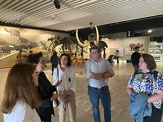
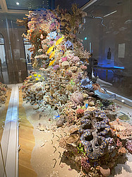
Hirnforschung meets Senckenberg
The Senckenberg Museum of Nature in Frankfurt is a great place to learn more about evolution, be fascinated by the combination of art and science, and discover that Frankfurt has its own little version of the Great Barrier Reef. The director, Professor Dr. Brigitte Franzen, gave us a great tour – thank you very much!
New release of the Julich-Brain Atlas adds 52 new maps
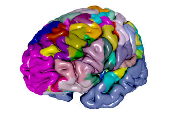
Release 3.1 of the Julich Brain Atlas has been published and can be freely downloaded through the EBRAINS research infrastructure. The updated brain atlas now gives online access to 52 new probability maps of cortical and subcortical structures in a three-dimensional reference space.
The Julich-Brain Atlas (Amunts et al. Science 2020) contains cytoarchitectonic maps of 227 areas of the human brain including cortical areas and subcortical nuclei. Based on differences in distribution, density and morphology of cells in a three-dimensional space it contains probabilistic maps that reflect the variability between individual brains. It represents the most comprehensive and complete microstructural map of the human brain to date.
The Julich Brain is the foundation of the Multilevel Human Brain Atlas on EBRAINS, which integrates neuroanatomical features with complementary maps of the molecular architecture, function and connectivity across multiple scales and is openly available to the research community. In this way the atlas serves as a basis for spatially aligning and annotating data and knowledge from different levels of brain organisation and as a powerful tool to help researchers and clinicians better interpret images of individual brains.
The Julich-Brain Atlas is a living atlas that is consistently being expanded and refined. Special features in Release 3.1 include the completely mapped thalamus with all its subnuclei, which are included in the Julich Brain Atlas for the first time – a result of a collaboration with Harry Uylings from Amsterdam.
All maps are available in commonly used reference spaces Colin27, MNI152 and fsaverage, which means that both a volume-based version with cortical and subcortical areas and a surface-based version with cortical areas are provided. Furthermore, rich meta-data is included to enable the provision of data via the EBRAINS infrastructure according to the FAIR principles.
Data in the EBRAINS KnowledgeGraph:
Amunts, K., Mohlberg, H., Bludau, S., Caspers, S., Lewis, L. B., Eickhoff, S. B., & Pieperhoff, P. Julich-Brain Atlas, cytoarchitectonic maps (v3.1) [Data set].
Paper reference:
Amunts K, Mohlberg H, Bludau S, Zilles K. Julich-Brain: A 3D probabilistic atlas of the human brain's cytoarchitecture. Science. 2020 Aug 21;369(6506):988-992. doi: 10.1126/science.abb4588.
Action Day at Green Zoo Wuppertal, May 25th, 2024
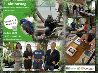
Once again, we took part in the 2nd action day of our cooperation partner ‘The Green Zoo’ Wuppertal together with Prof. Dr. Markus Axer (Bergische Universität Wuppertal/FZ Jülich). Guided by its motto ‘Nature protection, species protection, and climate protection’ and accompanied by curious glances of the male gorilla, a silverback named ‘Vimoto’, we presented rare exhibits from our institute and archive as well as many hands-on activities in the monkey house of Zoo Wuppertal. Among others, a brain cast of ‘Bobby’, the first gorilla of the Berlin Zoological Garden (1928) could be admired and touched. For our visitors - young & older ones – we offered a quiz which guided them through the various stations of our exhibition stand. Our visitors could learn about modern physical methods, such as 3D polarization microscopy, but also how cognitive abilities and skills are interconnected with brain organization. For this, brain slices were rotated on a hand polariser and with that changing connections in the brain could be observed by our visitors themselves. They could also search for the rat barrel cortex via a transmitted light microscope. In other challenges, our visitors could compete with pigeons and should distinguish between Monet and Manet. Or different brains should be sorted by their brain size to the appropriate species of songbirds and parrots, to get an understanding that enormous brain abilities can be developed despite having a small brain. Solving that quiz was really hard work, but everyone who finally handed in the completed quiz sheet was rewarded - this year with gummy bears, pens, and paper. With great joy, we have been supported by our student Lena Koukal (BSc Natural Sciences) and PostDoc Dennis Scheidt (FZ Jülich) this year. What else can we say? It was another great day, with many interested visitors of all ages. We hope we can pass on our enthusiasm to many of them. We want to thank the whole Green Zoo Wuppertal team for the great organization and implementation, especially our cooperation partner Dr. Dominik Fischer, who already invited us for the action day next year! (Source: CH)
Master’s program Translational Neuroscience at HHU Düsseldorf
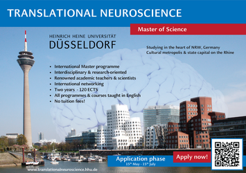
The application process for our master's program Translational Neuroscience will start on 15th May 2024. The deadline will end on 15th July 2024.
@international students: Please notice that you have to hand in a VPD (preliminary review documentation) this year. You have to apply on the website of uni-assist for it. https://www.uni-assist.de/en/tools/uni-assist-universities/detail/hochschule/123/
Further information can be found here: https://www.translationalneuroscience.hhu.de/
We are looking forward to your application!
BigBrain: A 3D atlas of the human brain and what it can do for medicine
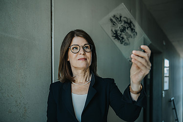
Prof. Dr. med. Katrin Amunts is Professor and Director of the C. and O. Vogt Institute of Brain Research at Heinrich-Heine-University Düsseldorf and Director of the Institute of Neuroscience and Medicine at Forschungszentrum Jülich. In the #ForscherinnenFreitag podcast, she talks about the digital atlas of the human brain, which she has been working on for 30 years. She provides insights into the development of brain research and her own career path. #ForscherinnenFreitag is an interview podcast from the #InnovativeWomen platform, which aims to make innovative women in science, business and society visible. www.innovative-frauen.de
The episode is available on the following page: https://www.innovative-frauen.de/podcast and on all common podcast platforms.
New study shows brain maps of the "red nucleus"
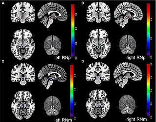
In a recent study, a German-American team of scientists has now revealed new insights into the evolutionary development of a brain structure that plays a crucial role in finely tuned and skillful hand movements. The researchers from C.&O. Vogt Institute for Brain Research at HHU Düsseldorf, Forschungszentrum Jülich and Stony Brook University in New York studied a brain structure called the “red nucleus” (lat. Nucleus ruber). This nucleus is an important element of our motor system, especially for dexterous hand movements.
However, many questions about the structural and functional evolution of this nucleus remain enigmatic. The brain researchers created cytoarchitectonic probabilistic maps of this nucleus that capture the variations in structure between brains. Furthermore, using brain sections from the large brain collection in Düsseldorf, the authors could perform a phylogenetic analysis uncovering the evolutionary development of this structure in primates. They could show that two different subdivisions of the red nucleus underwent distinct evolutionary trajectories. While the phylogenetically older part has become smaller in humans and apes, the second part has actually grown in proportion to brain size in all primates, including humans. This is why the red nucleus in humans and great apes, in contrast to other primates, consists of a large second part and a vestigial other part.
These important insights into the structural evolution of the red nucleus spark new hypotheses about its functional specialization, e. g. whether these changes may play a positive role in neurodegenerative diseases. The new maps of the red nucleus can be utilized for future research that will further advance our understanding of the role of this structure in motor and cognitive
Stacho M, Häusler AN, Brandstetter A, Iannilli F, Mohlberg H, Schiffer C, Smaers JB and Amunts K (2024) Phylogenetic reduction of the magnocellular red nucleus in primates and inter-subject variability in humans. Front. Neuroanat. 18:1331305. https://doi.org/10.3389/fnana.2024.1331305
New brain maps on EBRAINS show organizational principles of the prefrontal cortex
German researchers have published new brain maps on EBRAINS, identifying five new areas within the dorsolateral prefrontal cortex (DLPFC), that plays a crucial role in executive functions, including working memory, decision-making, and attention. The new study introduces 3D maps of cytoarchitectonic regions, enhancing our understanding of the prefrontal cortex's complexity.
The study is an important step toward deciphering the structural-functional organization of the human prefrontal cortex. By analyzing series of histological sections and employing state-of-the-art statistical techniques, Bruno & Lothmann et al. identified and mapped five new areas within the DLPFC, and revealed their variations across brains. The three-dimensional maps will be part of the next release of the Julich Brain Atlas, which is freely accessible via the EBRAINS research infrastructure.
Original publication: Bruno, A., Lothmann, K., Bludau, S., Mohlberg, H., Amunts, K., New organizational principles and 3D cytoarchitectonic maps of the dorsolateral prefrontal cortex in the human brain. Front. Neuroimaging, 3 (2024), DOI: https://doi.org/10.3389/fnimg.2024.1339244
Registration and call for abstracts to the Helmholtz AI Conference 2024 is OPEN!

Join us this June 12 - 14, 2024 at the CCD Congress Center Düsseldorf in Düsseldorf, Germany, and unravel the capabilities of AI to reshape scientific research through more than 30 in-depth sessions, special keynote talks, a poster showcase and plenty of networking opportunities. Secure your spot and register at our conference website!
Engage with method and domain experts and world-leading scientists in AI research; we have prepared a myriad of opportunities for connecting with others, from our official conference dinner reception to a brand new edition of the beloved Unconference of last year, a world café and a whole day of lab touring, workshops and hackathons at the Prologue Day on June 11 at Forschungszentrum Jülich! See more about our program and satellite events on the conference website.
Congress "Evolutionary Roots of Human Brain Diseases"

From February 22 to 24, 2024, the Congress "Evolutionary Roots of Human Brain Diseases" will take place at the Hotel Légère in Muensbach, Luxembourg. The Scientific Committee consists of Prof. Katrin Amunts, Prof. Nico J. Diederich, Prof. Martin Brüne and Prof. Christopher G. Goetz.
Further information on registration and the program can be found on the website: https://www.chl.lu/fr/congress-evolutionary-roots-human-brain-diseases
New year and new colleagues at the Institute of Brain Research
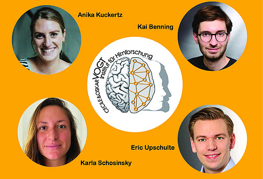
We are pleased to announce that Anika Kuckertz, Karla Schosinsky, Kai Benning, and Eric Upschulte have joined our team and are pursuing new research projects. Welcome to the team! We wish you a great start and are looking forward to fruitful collaborations.
“Impressive research results” - external review panel evaluates final results of Human Brain Project

From 21-24 November, the final Human Brain Project (HBP) review was held in Brussels during which members of the HBP consortium presented the final project results to a panel of external scientific experts. The scope of this review was the final phase of the HBP, which ended in September 2023. Results were presented in detailed documentation and presentations and followed by extensive Q&As.
The expert panel drew a strongly positive conclusion when announcing the preliminary results of their review of the HBP to the consortium and the European Commission (EC) on Friday.
The panel provided overall preliminary comments that are reproduced below. The reviewers emphasized the impressive results of the project and that the legacy of the HBP can be “fundamental in a new phase of neuroscience”, as the project laid the foundations to enable research collaboration on a very large scale, with lasting implications for innovation and the understanding of the brain. The in-depth written review report will now be finalized and is expected to be provided to the European Commission in early 2024.
The full preliminary statement of the review panel:
“The HBP has achieved impressive research results and delivered an open research infrastructure positioned to have a transformative impact in Europe and beyond. Already today, the EBRAINS infrastructure empowers new applications in brain health, and brain derived technologies. The HBP has established a new paradigm of digital neuroscience and a new interdisciplinary culture of collaboration.
Particular highlight achievements include leading digital brain atlases, advanced brain simulation platforms across scales, application of cognitive modelling and personalized medicine, as well as outstanding advances in neuromorphic computing and neuro inspired robotics and AI.
In light of these achievements, the HBP catalyzed highly interdisciplinary research at the scale enhancing our understanding of the brain.
The legacy of the HBP can be fundamental in a new phase of neuroscience.”
Media contact
Original article can be found here.
Katrin Amunts honored with international Justine and Yves Sergent Award
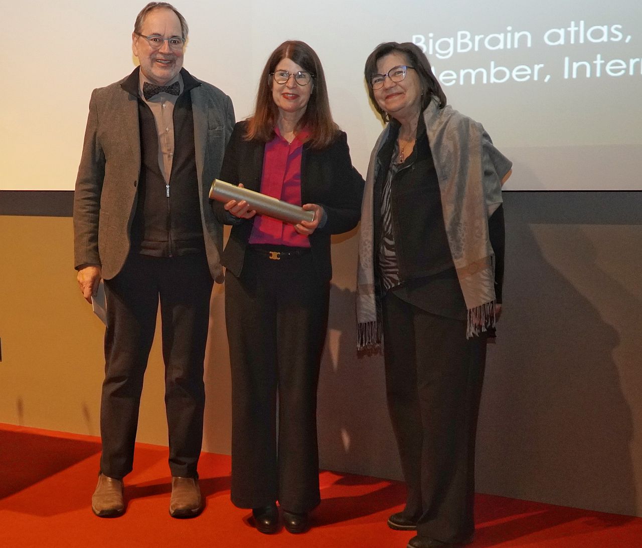
Copyright: University of Montreal
Prof. Katrin Amunts was honored with the international Justine and Yves Sergent Award 2023. The award is presented to internationally renowned female researchers for outstanding achievements in cognitive neuroscience and brain imaging. The award ceremony took place during a conference at the University of Montreal (Canada) on Friday, December 8. Katrin Amunts gave a keynote speech on the topic of "Human Brain Atlas - Mapping the human brain to better understand its functions".
Please find here further information.
Katrin Amunts Appointed to Leopoldina
A special honour for an outstanding brain researcher: Prof. Katrin Amunts was appointed to the German National Academy of Sciences Leopoldina and will be active in the Psychology and Cognitive Sciences Section. Leopoldina is the oldest continuously existing academy of natural sciences and medicine in the world. Leopoldina selects its members among scientists who have distinguished themselves through their outstanding scientific achievements.
Please find here further information.
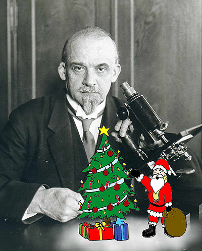
On behalf of the entire team, the Cécile & Oskar Vogt Institute of Brain Research wishes a Merry Christmas and a Happy New Year 2024!
Woman in MICCAI (WiM)
Nataliia Fedorchenko and Ahmed Nebli (INM-1, Research Centre Juelich) won the third prize for their video about Women in MICCAI (WiM), Inspirational Leadership Legacy of the Medical Image Computing and Computer Assisted Intervention Society (MICCAI).
Congratulations to this award!
Please find here the announcement and the video.
Save the Date: Helmholtz AI Conference 2024
12 -14 June 2024, Düsseldorf
Save the date for 12 - 14 June, 2024 in Düsseldorf!
AI has the power to accelerate discoveries and aid us in building a better tomorrow faster – as long as we share the invaluable knowledge we possess with others. Building upon the success of our last conference in 2023, this next edition is titled "Helmholtz AI Conference 2024: AI for Science." The event is scheduled to take place from June 12th to 14th at the CCD Congress Center Düsseldorf in Düsseldorf, Germany.
Please find here further information.
Leanne Demay is a visiting scientist at the C.&O.Vogt Institute of Brain Research

Leanne Demay is a PhD candidate at Stony Brook University New York in the Department of Anthropology and currently a visiting researcher at the C.&O. Vogt Institute of Brain Research. Her dissertation is a comparative analysis of the cerebellar lobules of primates. Here on site, she has the opportunity to use our extensive histological brain slice collection to analyze the interspecific variables based on high-resolution scans of the cerebellar lobules of various primate species. We wish Leanne a good start to her project and a pleasant stay with us in Düsseldorf.
Further team growth at the Institute of Brain Research
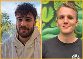
We are very happy that Rouven Fleischmann and Simon Werker have joined our team. A warm welcome! We wish you all the best for your start and are looking forward to working with you. Rouven will support us as a biological-technical assistant in the lab and Simon will help out as a student assistant.
The Human Brain Project ends: What has been achieved
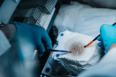
On September 30th, the Human Brain Project (HBP) formally completes its 10-year runtime as an EU-funded FET Flagship. The project has pioneered digital neuroscience, a new approach to studying the brain based on multidisciplinary collaborations and high-performance computing. The HBP will continue to have an impact on neuroscience for many years through the EBRAINS research infrastructure and a new way of collaborative work in the field.
Read the further article here.
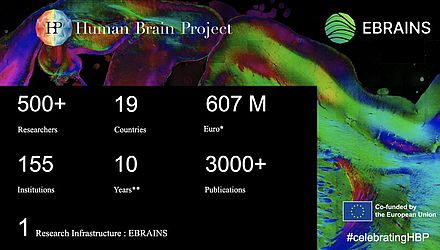
After 10 years , the EU Flagship Human Brain Project will come to an end on 30 September. We’re very proud to have been a partner of the project.
Learn more about the project’s achievements here https://www.humanbrainproject.eu/ #celebratingHBP
Human Brain Project celebrates successful conclusion
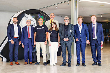
Copyright: Forschungszentrum Jülich
The EU-funded Human Brain Project (HBP) comes to an end in September and celebrates its successful conclusion today with a scientific symposium at Forschungszentrum Jülich (FZJ). The HBP was one of the first flagship projects and, with 155 cooperating institutions from 19 countries and a total budget of 607 million euros, one of the largest research projects in Europe. Forschungszentrum Jülich, with its world-leading brain research institute and the Jülich Supercomputing Centre, played an important role in the ten-year project.
Read more here.
Welcome to the team!
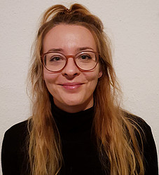
We are very happy to welcome Dr. Lucija Rapan as a new research associate at the Cécile & Oskar Vogt Institute of Brain Research.
Lucija will support our research and teaching activities from now on. Her research focus is the characterization of the cytoarchitecture and receptor architecture of the posterior cingulate and retrosplenial cortex of humans and macaques.
We wish you a good start, Lucija!
Max Planck School of Cognition - application portal is open now
The application portal is open from September 1st 2023 until December 1st 2023 for intake 2024!
Please find further information on their website: https://cognition.maxplanckschools.org/en/application
Action Day Wuppertal Zoo from May 13, 2023
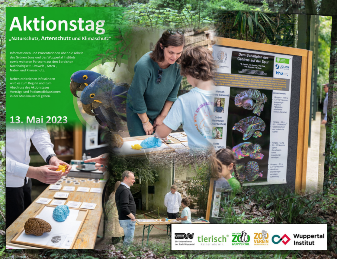
Together with Prof. Dr. Markus Axer (Bergische Universität Wuppertal/FZ Jülich) we participated in the action day "Nature Protection, Species Protection and Climate Protection" of our cooperation partner "The Green Zoo Wuppertal". We presented the brain diversity of different species all visualized by 3D polarization microscopy. Besides, we showed impressive specimens from our brain and brain slice collection - from orangutans to crocodiles. Our young as well as adult visitors enjoyed our little memory game "Which brain belongs to which species?". Our visitors themselves could also experience the physics behind that 3D-PLI method by using our "hand polarimeter" and could try themselves in further experiments. Our stickers were very popular and were taken home by many visitors. We were supported by our students Alina Hansen (M.Sc. Psychology) and Kevin Flink (B.Sc. Medical Physics). We would like to thank the whole team of the Wuppertal Zoo and especially our cooperation partner Dr. Dominik Fischer for this successful event! Next year we will be back to inspire again many visitors and future scientists, and with that, making a lasting contribution to our planet. Author: PD Dr. Christina Herold, Source photos: Wuppertal Institute / L.Schenk
Invitation to the 1st EBRAINS National Node Germany Workshop
We would like to kindly invite you to participate in the 1st EBRAINS National Node Germany Workshop which will take place on September 26, 2023 in Berlin from 09:00-13:30 CET.
Guest Speakers:
- Dr. Seán Froudist-Walsh, University of Bristol
- Prof. Dr. Thomas Wachtler, Ludwig-Maximilians-University Munich
- Prof. Dr. Lena Oden, FernUni Hagen & FZ Jülich
- Prof. Dr. Bryan Strange, Technical University Madrid
- Dr. Andreas Rowald, Friedrich Alexander University Erlangen-Nürnberg
- Prof. Dr. Petra Ritter, Charité – Universitätsmedizin Berlin
- Prof. Dr. Thanos Manos, Cergy Paris University
For further details and for registration please browse to our event website
The EBRAINS National Node Germany (NNG) invites to the 1st EBRAINS NNG Workshop as a back-to-back event of the Bernstein Conference 2023 in Berlin, Germany. You will hear about the EBRAINS RI, its usage, an Expert Discussion on “Digital tools to bridge the gap between experimental and computational neuroscience” and will have the opportunity to network during our NNG lunch.
EBRAINS is a unique digital Research Infrastructure (RI), created by the EU-funded Human Brain Project. It is an open platform providing an extensive range of data and tools to enhance brain-related research. EBRAINS is a pan-European distributed network of services, that will in the future be organized through National Nodes.
The EBRAINS NNG currently encompasses about 40 universities and research institutions across Germany joining forces to develop a common scientific and technical strategy, addressing the major scientific challenges of our time. The EBRAINS NNG partners offer unique services, including science liaison activities, for basic neuroscience, medical applications, and industry in the fields of:
- High-Performance Computing
- Neuromorphic Computing
- Artificial Intelligence
- Simulation
- Robotics
- Information privacy and security
Programme Committee:
Nicola Palomero-Gallagher, Fabrice Morin, Francesca Cavallaro, Björn Kindler, Sandra Diaz, Maren Frings
In case of further questions please don’t hesitate to contact ebrains-nng@fz-juelich.de
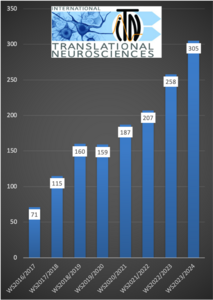
Our Master's program in Translational Neuroscience is becoming more and more visible and popular every year - both nationally and internationally. More and more young scientists are interested in obtaining a Master's degree in neuroscience and are applying for our program here at Heinrich-Heine-University Düsseldorf. This year we have already set a new record and exceeded 300 applications. That is +18.2% more applications than last year and even 4 times more than in 2016 - when our program was launched for the very first time. We wish all applicants good luck and every success! We will personally welcome 20 of you here in Düsseldorf for the start of the winter semester 2023/24 in October. And, you can still apply!
The application deadline is on Saturday, the 15th of July 2023, 23:59 o’clock:
HBP researchers identify three new human brain areas involved in sexual sensation, motor coordination, and music processing
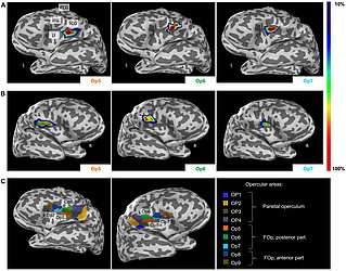
HBP researchers from Germany performed detailed cytoarchitectonic mapping of distinct areas in a human cortical region called frontal operculum and, using connectivity modelling, linked the areas to a variety of different functions including sexual sensation, muscle coordination as well as music and language processing.
You can find the complete article here.
Reference
Nina Unger, Martina Haeck, Simon B. Eickhoff, Julia A. Camilleri, Timo Dickscheid, Hartmut Mohlberg, Sebastian Bludau, Svenja Caspers and Katrin Amunts. Cytoarchitectonic mapping of the human frontal operculum-New correlates for a variety of brain functions. Front. Hum. Neurosci., 28 June 2023 – Sec. Speech and Language. Volume 17 – 2023. doi : 10.3389/fnhum.2023.1087026
Two interesting studies published in Communications Biology
Our colleague Felix Ströckens has recently published two highly interesting research studies in the journal communications biology.
The study on hemispheric asymmetries and brain size in mammals was done in collaboration with the Medical School and the Institute for Cognitive and Affective Neuroscience in Hamburg and the Institute for Cognitive Neuroscience at Ruhr University Bochum. If you want to know more about why larger-brained species tend to evolve into more right-lateralized individuals, we highly recommend this article.
Ocklenburg S, El Basbasse Y, Ströckens F, Müller-Alcazar A. Hemispheric asymmetries and brain size in mammals. Commun Biol 2023; 6(521), https://doi.org/10.1038/s42003-023-04894-z
The second study already has the delightful title ‚From Fossils to Mind‘. Fossil endoclasts preserved features of brains from the past (such as size, shape, vasculature, and gyrification) that can be used today to gain new scientific insights. Interesting facts about paleoneurology and how interdisciplinary techniques have made it possible to draw conclusions about the evolution and physiology of the brains of extinct species and how to study the dead within the living can be read in this article.
de Sousa AA, Beaudet A, Calvey T, Bardo A, Benoit J, Charvet CJ, Dehay C, Gómez-Robles A, Gunz P, Heuer K, van den Heuvel MP, Hurst S, Lauters P, Reed D, Salagnon M, Sherwood CC, Ströckens F, Tawane M, Todorov OS, Toro R, Wei Y. From fossils to mind (2023), Commun Biol 2023; 6(1), https://doi.org/10.1038/s42003-023-04803-4
HFA-Symposium 2023
Light in Biology - Photosynthesis, Visual Processes and Neuronal Applications
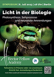
All life processes on our earth depend on the energy of sunlight. Light enables humans and animals to perceive their environment in colour and to orient themselves. But not only that - it also regulates their day-night rhythm and all related activities. In plants, light forms the basis for growth and development.
What role does light play in photosynthesis, the process that sustains life on our planet? How do animals perceive the world depending on the light environment in which they live? How can light control nerve cells and thus help us better understand how our memory works?
The event continues the series of Germany-wide symposia of the Hector Fellow Academy (HFA). Here, renowned experts present current research topics in a generally understandable way and discuss visions for the future. The HFA is a young science academy that promotes interdisciplinary cutting-edge research in MINT (mathematics, informatics, natural science and technology), psychology and medicine.
Here you can find further information about the programme
Location:
Langenbeck-Virchow-Haus, Luisenstraße 58/59, 10117 Berlin
Online participation:
You will receive the access data by e-mail.
Admission is free. The event will be held in German/English. Simultaneous translation will be provided.
Register now for the symposium "Light in Biology"! https://express.converia.de/frontend/index.php?sub=1188
Vogt-Archive visited by Augustana Neurophilosophy Summer School
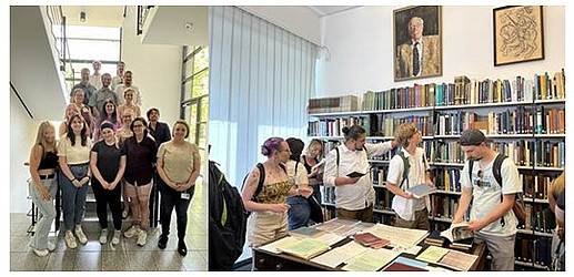
On 20. June 2023, the Vogt archive welcomed guests from Augustana College, Illinois, USA. The course of twelve students, led by Philosopher Heidi Storl and Neuroscientist Ian Harrington, also visited Forschungszentrum Jülich, in particular the Institute of Neuroscience and Medicine and the Jülich Supercomputing Center.
Introductions to the two scientific archives were given by institute director Katrin Amunts and archivist Caroline Laperrouze (C. u. O. Vogt Institute for Brain Science at UKD) as well as Fabio de Sio and Ulrich Koppitz (Eccles-Archive, Institute for the History, Philosophy and Ethics of Medicine at HHU).
Thank you for the visit and the interesting discussions!
Human Brain Project study offers insights into neurotransmitter receptor organisation
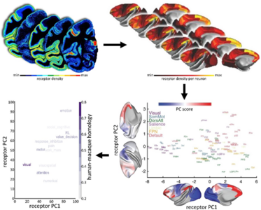
A key challenge in neuroscience is to understand how the brain can adapt to a changing world, even with a relatively static anatomy. The way the brain’s areas are structurally and functionally related to each other – its connectivity – is a key component. In order to explain its dynamics and functions, we also need to add another piece to the puzzle: receptors. Now, a new mapping by Human Brain Project (HBP) researchers from the Forschungszentrum Jülich (Germany) and Heinrich-Heine-University Düsseldorf (Germany), in collaboration with scientists from the University of Bristol (UK), New York University (USA), Child Mind Institute (USA), and University of Paris Cité (France) made advances on our understanding of the distribution of receptors across the brain.
The findings were published in Nature Neuroscience, and the data is now freely available to the neuroscientific community via the HBP’s EBRAINS infrastructure.
The HBP team used autoradiography to analyse the density of receptors for neurotransmitters on very thin in vitro brain sections. They measured the density of 14 neurotransmitter receptor types in 109 areas of the macaque cortex and this data was integrated with multiple structural parameters into neuroimaging templates.
Neurotransmitter receptors
Receptors are key molecules in signal transmission in the brain. Within a neuron, information transmission occurs via electric signals along the axon. But transfer of information between neurons usually requires the release of molecules called neurotransmitters into the extracellular space and their binding to receptors on the target neuron.
The HBP researchers have uncovered a primary and a secondary gradient of receptor expression per neuron. In other words, they mapped receptor densities across the cortex and were able to identify two main arrangements, shedding light on the links between molecular and neuron organization of the cortex. “These two major axes of receptor organisation in the macaque cortex align with two different functional systems, namely the sensory-cognitive and the external-internal cognition networks. This is the first time that such an association has been described,” explains Nicola Palomero-Gallagher, researcher at the Forschungszentrum Jülich and senior author of the paper.
Integrating maps
In their study, the researchers integrated the new neurotransmitter receptor data with multiple layers of anatomical and functional data onto a common cortical space within the cortical surface of Yerkes19, a frequently used non-human primate template. Few studies so far had integrated in vitro anatomy and in vivo imaging of the macaque brain. Creating openly-accessible maps of receptor expression across the cortex that integrate neuroimaging data, such as what was done by the HBP team, could speed up translation across species.
“It is being made freely available to the neuroscientific community so that they can be used by other computational neuroscientists aiming to create other biologically informed models,” Palomero-Gallagher says. Part of the data generated for this study has already been implemented in a computational model of how dopamine gates information into the frontoparietal working-memory network.
Text by Helen Mendes
Original Publication:
Gradients of neurotransmitter receptor expression in the macaque cortex
Sean Froudist-Walsh, Ting Xu, Meiqi Niu, Lucija Rapan, Ling Zhao, Daniel S. Margulies, Karl Zilles, Xiao-Jing Wang, Nicola Palomero-Gallagher. Nature Neuroscience. 19 June 2023. https://www.nature.com/articles/s41593-023-01351-2
DOI 10.1038/s41593-023-01351-2
Congratulations on your doctorate, Dr. Bruno!
Today our doctoral student Ariane Bruno successfully defended her dissertation at the Medical Faculty of Heinrich Heine University Düsseldorf. An incredible achievement, a lot of diligence, and the result of hard work.
We are very proud of you!
Congrats from the whole team of the C.&O. Vogt Institute of Brain Research!
Join us for the concluding event of the Human Brain Project - Pioneering digital brain research
12-13 September 2023 | Forschungszentrum Julich, Germany
Scientific Symposium | lab tours | exhibition | reception
From September 12 - 13, 2023, the Human Brain Project will celebrate its successful conclusion with a scientific symposium at Forschungszentrum Julich. In addition to the international project partners, representatives from politics and the media are cordially invited to attend.
In short presentations, researchers from the Human Brain Project will highlight the project’s achievements. The symposium will be accompanied by scientific exhibits, an impressive picture gallery and hands on trainings. In guided tours guests can explore the laboratories and facilities of Forschungszentrum Julich and get insights into the practical work behind the Human Brain Project.
For more information, please click here: https://www.humanbrainproject.eu/en/hbp-final-event/
Congratulations to our PhD student Serap Kurutas on her doctorate! Yesterday, Serap successfully defended her dissertation at the Medical Faculty of the Heinrich Heine University Düsseldorf. Prof. Dr. Katrin Amunts, PD Dr. Christina Herold and PD Dr. Julia Mehlhorn and both teams from the Cécile & Oskar Vogt Institute of Brain Research and Anatomy I at University Hospital Düsseldorf are very proud of you, Doctor Kurutas! Congrats from all of us!
Master's Program Translational Neuroscience
Our Master's program Translational Neuroscience at HHU Düsseldorf is open for applications!
We are looking forward to your application on https://digstu.hhu.de/qisserver/pages/cs/sys/portal/hisinoneStartPage.faces
Application Deadline on 15th July 2023
Further infomation regarding the program can be found on our website https://www.translationalneuroscience.hhu.de/
Research subjects wanted for Puppilox study
We are looking for subjects for the Pupillox study - a computer-based behavioral study to investigate pupil dilation during transcutaneous vagus nerve stimulation.
The aim of the study is to investigate whether transcutaneous ("through the skin") vagus nerve stimulation (tVNS) leads to changes in pupil diameter. tVNS is a safe, non-invasive procedure in which the vagus nerve is stimulated through the skin of the left ear. It involves placing two small electrodes in two locations on your left ear, through which small amounts of electrical current flow. The electrode gel is applied to the inner ear only.
Further information about the study and registration can be found here.
Research ubjects wanted to study Ataxia & Parkinson's disease
The Clinical Neuroanatomy group of the Institute of Neuroscience and Medicine at Research Centre Jülich is looking for patients as well as healthy control subjects for research on Ataxia & Parkinson's Syndrome.
If you are interested in one of the mentioned studies, please contact the respective study contact person or send an email to m.minnerop@fz-juelich.de. Please include the study name, the study you are interested in, your name and phone number. You will then be called back promptly by the respective contact person.
Further information about the study can be found here.
The gap in our team is huge. The memory of you remains alive in our hearts.

Ulrich Opfermann-Emmerich
* 9th January 1961 ✝ 24th December 2022
We are deeply saddened by the death of our dear colleague who died much too soon. With Ulrich Opfermann, we lose an always helpful, friendly and committed employee. For more than 25 years, Uli was not only a medical-technical employee in our laboratory, but also responsible for the care of the Vogt archive and our collections, he was our house and court photographer and the good soul of the institute.
Our sincere condolences and deepest sympathy are with Uli's family.
We will always keep you in good memory and never forget your words: "Please smile".
Your colleagues of the Cécile & Oskar Vogt Institute of Brain Research
For the reception in the new Vogt Archive in the Himmelgeisterstraße on the 10th of January 2023, employees of the C.&O. Vogt Institute of Brain Research, representatives of the Medical Faculty, the Institute of the History, Philosophy and Ethics of Medicine, the Gesellschaft von Freunden und Förderern der Heinrich-Heine-Universität e.V as well as the members of the Board of Trustees and donors of the Cécile and Oskar Vogt Foundation came together. It was the kick-off for the digitization and relocation project of the estate and brain section collection of the research couple Cécile and Oskar Vogt. Many thanks to all participants who made this evening an impressive event. Especially to our speakers for their warm greetings, in particular to our Institute Director Prof. Dr. Amunts, to Prof. Dr. Nikolai Klöcker, Dean of the Medical Faculty, to Prof. Dr. Heiner Fangerau, Vice Dean for Strategic Development, to Mr. Eduard Dörrenberg, President of the Gesellschaft von Freunden und Förderern der Heinrich-Heine-Universität e.V. (GFFU) and to our guest of honor Prof. Dr. Rita Süssmuth. In the upcoming months, the Vogt Collection will be successively moved to its new location, digitized. Thus much easier access to this historical research material will be provided. It will help to complete our brain mapping and integrate the research results of more than six decades from the late 19th century into the modern brain atlases of the 21st century.
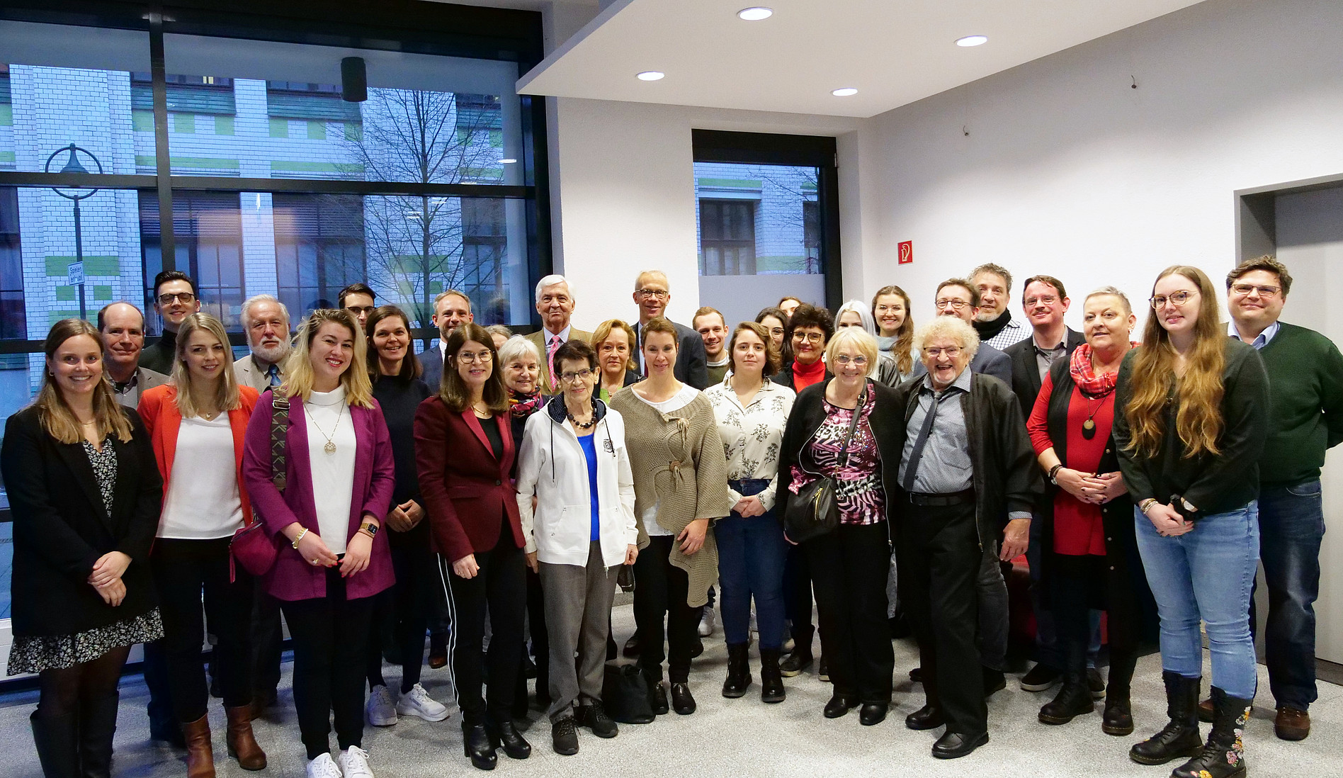
Registration for the Human Brain Project Summit 2023 is open
The Human Brain Project (HBP) is delighted to announce that registration for the HBP Summit 2023 is open. The event will take place at the Palais du Pharo in Marseille, France, from March 28-31, 2023.
Registration
Register before the early bird deadline on 2 February to benefit from a reduced registration fee! Click here to register.
Abstract submission
Would you like to present your research at the HBP Summit? Make sure to submit your poster abstract! Two winners will have the opportunity to present their research to the audience in a 10-minute plenary talk. Submit your abstract before 2 February here.
Satellite events
A selection of satellite events - covering topics such as simulation, atlasing, and data analytics - will take place on 27 March 2023 at the Palais du Pharo as part of the Human Brain Project Summit 2023. You can find a preliminary list of satellite events in the programme here. Please note that you will have to register separately for each satellite event you wish to attend.
Please find here further information.
Unique brain collection to be digitized: Kick-off at the Cécile and Oskar Vogt Archive
Oskar and Cécile Vogt were early pioneers in mapping the brain. In the six decades of their work, they made significant contributions to the understanding of brain organization and to the development of brain research as a science. The C.&O. Vogt Archive contains the extensive estate of the research couple.
To mark the start of the digitization and relocation of the Vogt Archive, brain researcher and institute director Prof. Katrin Amunts invites to a kick-off meeting in the new building in Düsseldorf's Himmelgeisterstraße. Invited are the staff of the C.&O. Vogt Institute of Brain Research, representatives of the Medical Faculty and the Institute of History, Philiosophy and Ethics of Medicine, as well as the Gesellschaft von Freunden und Förderern der Heinrich-Heine-Universität e.V., and the members of the Board of Trustees and donors of the Cécile and Oskar Vogt Foundation.
The Cécile and Oskar Vogt Institute of Brain Research at UKD and HHU is in the tradition of the research conducted by the Vogts and focuses on the question of the basic organizational principles and functions of the human brain, in particular the cerebral cortex and its dependence on various internal and external influences.
Date:
Tuesday, January 10, 2023 at 4 PM
Location: Himmelgeister Straße 103-105 in 40225 Düsseldorf
Contact: Caroline Laperrouze, scientific archivist (calap101@hhu.de)
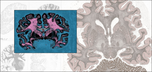
A successful research year 2022 comes to an end for our scientists at the Cécile & Oskar Vogt Institute of Brain Research at University Hospital Düsseldorf and Institute for Neurosciences and Medicine (INM-1) at Forschungszentrum Jülich. Many thanks to everyone for the first-class research projects, collaborations, and publications in the renowned scientific journals 2022.
We wish you a Merry Christmas and a Happy New Year 2023.
Nataliia Fedorchenko awarded place in prestigious Max Planck School of Cognition

Ukrainian brain researcher Nataliia Fedorchenko has been accepted as an associated PhD candidate at the Max Planck School of Cognition (MPSCog). She will carry out her PhD under the supervision of MPSCog faculty member Katrin Amunts at the Cécile and Oskar Vogt Institute of Brain Research at Heinrich Heine University Düsseldorf.
Fedorchenko joined Amunts’ team earlier this year after fleeing Kyiv, where she had recently started a PhD focused on stroke after Covid-19 infections. The trained neurologist has a special interest in stroke, speech deficits and language cognition.
In Jülich and Düsseldorf, Fedorchenko currently works within the scientific coordination team of the Human Brain Project, where she is mainly responsible for the position paper ‘The coming decade of digital brain research – A vision for neuroscience at the interaction of technology and computing’. She is also involved in the digitalization of the Vogt brain collection at the Cecile & Oskar Vogt Institute.
In January 2023, Fedorchenko will commence her new PhD project studying the Broca's region of the human brain, which is involved in language processing and plays a major role in the clinical context, e.g., in aphasia.
To unveil the connectivity of different areas within the brain region, Fedorchenko will employ an advanced imaging technique called 3D Polarized Light Imaging, which has been developed by Amunts’ team. Her aim is to achieve novel insights into the region’s organization that will eventually advance clinical applications.
Fedorchenko is one of four associated candidates who have been accepted this year to the highly competitive programme of the Max Planck School of Cognition. PhD students of the graduate school conduct research at world-renowned research institutions in Germany, the Netherlands and the UK. As an associated PhD candidate, Fedorchenko will join the Max Planck School for three years.
Fedorchenko is particularly excited about the interdisciplinary approach and the international network of brilliant scientists that the Max Planck School is offering. “Being part of the Max Planck School of Cognition is a unique opportunity to have access to high-level researchers and receive valuable feedback from other international students,” she says.
Read more about Nataliia Fedorchenko in the Helmholtz news:
https://www.helmholtz.de/en/newsroom/article/leave-of-absence/
Success in international AI-competition
The annual NeurIPS conference (Conference on Neural Information Processing Systems) is considered the world's most important forum for artificial intelligence, with participants from all over the world. Every year, competitions are held as part of the conference to select the best AI developments for various use cases.
In the category "Machine Learning for Physical and Life Sciences", Eric Upschulte, a member of the Big Data Analytics group at INM-1, placed one of the top spots. He reached the top ten among 443 participants in the "Cell Segmentation Competition". The researcher will now present his results at the conference in New Orleans, USA.
For the competition, participants submitted self-developed AI algorithms that automatically recognise cells in microscopy images. Such methods are becoming increasingly important in order to be able to deal efficiently with large data sets from research. At INM-1, the algorithms contribute to mapping the brain by quickly recognising and labelling individual neurons in large high-resolution scans.
The 36th Conference on Neural Information Processing Systems (NeurIPS) will take place from 28 November to 09 December 2022. More information about the competitions:
https://neurips.cc/Conferences/2022/CompetitionTrack
Multilevel brain atlases provide tools for better diagnosis
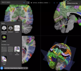
The multilevel Julich Brain Atlas developed by researchers in the Human Brain Project, could help in studying psychiatric and aging disorders by correlating brain networks with their underlying anatomical structure. By mapping microarchitecture with unprecedented levels of detail, the atlas allows for better understanding of brain connectivity and function. Researchers of the HBP have provided an overview of the Julich Brain Atlas in the journal Biological Psychiatry. The paper focuses on the cytoarchitecture and receptor architecture of the human brain, and how to apply the atlas in the field of psychiatric research.
Cytoarchitecture, the study of the distribution, density and morphology of cells in the nervous system, has a long standing history in brain mapping. Neuroscientists first noticed structural differences between areas of the cortex back in the late 1800s and started dividing it into distinct areas. The areas have been considered important correlates with brain function and dysfunction. In addition to the cellular architecture, the Julich Brain Atlas also includes maps of the distribution of receptors for the neurotransmitters which modulate brain activity. Neurotransmitter receptors differ not only between areas, but also between the different layers of an area and thus are closely related to its connectivity pattern, and relevant for its role in larger networks. Based on the data collected from post mortem brains, the atlas accounts for the naturally occurring variability between subjects by producing probabilistic maps in 3D spaces instead of the map of an individual brain only.
The Julich Brain Atlas is a “living” atlas that grows with new insights into the parcellation of the brain constantly being integrated. It is linked to other maps, e.g., coming from studies of fiber tracts in the living human brain. Such macroscopic and microscopic data are integrated in the HBP’s Multilevel Human Brain Atlas, which is openly accessible on the EBRAINS digital research infrastructure through the siibra software tool suite.
The researchers listed recent use cases of the tools in different peer-reviewed studies. Users can, for example, analyse and share high-resolution imaging data and compare it with fMRI datasets. They can look into the cytoarchitecture of a certain region and its connectivity, both within itself and with other regions. With a special tool called JuGEX, the maps can be linked to gene expression data from the Allen Brain Atlas, allowing deep multimodal investigations: For example, using the Julich Brain Atlas, researchers had identified new brain areas that play a role in major depressive disorder. Neuroimaging data from patients revealed area-specific changes in gray matter volume and activation. With JuGEX, these findings were further linked to local differences in the expression of several candidate genes for major depressive disorder. From large population studies, individual, personalised maps of aging or dysfunction can also be extracted to provide diagnostic tools for dementia.
Text by Roberto Inchingolo
Reference: Daniel Zachlod, Nicola Palomero-Gallagher, Timo Dickscheid, Katrin Amunts (2022). Mapping cyto- and receptor architectonics to understand brain function and connectivity. Biological Psychiatry, DOI: doi.org/10.1016/j.biopsych.2022.09.014
INM-1 contributes to National Research Data Infrastructure for Microscopy and Image Analysis

At the beginning of November, the Joint Science Conference (GWK) decided to include eight more consortia in a third round of funding for the National Research Data Infrastructure (NFDI). This means that the NFDI4BIOIMAGE consortium will also be funded for the next five years. At INM-1, Prof. Timo Dickscheid's research group "Big Data Analytics" is involved in the consortium. https://www.fz-juelich.de/en/inm/inm-1/research/big-data-analytics
The National Research Data Infrastructure NFDI is an initiative to establish an infrastructure framework for research data management in Germany. The aim is to develop reliable central standards and infrastructures for the storage, networking and use of data from science and research. This should enable data to be used across disciplinary and institutional boundaries.
The NFDI4BIOIMAGE initiative focuses on microscopy and biological image acquisition and processing ("bioimaging"). It is precisely in these areas that huge amounts of research data accumulate. For many disciplines in the life sciences that rely on bioimaging, adequate data management is indispensable, but it has not yet been sufficiently realised.
NFDI4BIOIMAGE aims to create solutions so that bioimaging data can be shared and reused across disciplinary boundaries. This should enable the full information content of the data to be exploited. They should also be available for re-analysis in order to gain new insights - possibly beyond the original research question.
A combination of micro and macro methods sheds new light on how different brain regions are connected
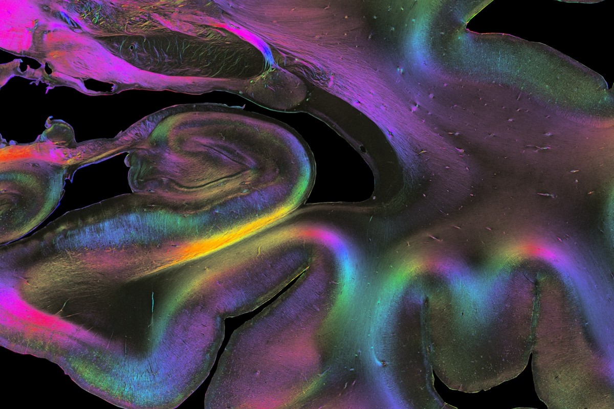
To understand how our brain works, there is no getting around investigating how different brain regions are connected with each other by nerve fibres. In the most recent issue of Science, researchers of the Human Brain Project (HBP) review the current state of the field, provide insights on how the brain’s connectome is structured on different spatial scales – from the molecular and cellular to the macro level – and evaluate existing methods and future requirements for understanding the connectome’s complex organisation.
Read the full article here.
Original publication:
Markus Axer & Katrin Amunts. Scale matter: The nested human connectome. Science, 3 Nov 2022. Vol 378, Issue 6619, pp. 500-504, DOI: 10.1126/science.abq2599
Atlasing in rodents:
In the same issue, HBP Infrastructure Director Jan Bjaalie (University of Oslo) and principal investigator Trygve B. Leergaard review brain data integration in rodent atlases:
Leergaard & Bjaalie. Atlas-based data integration for mapping the connections and architecture of the brain. Science, 3 Nov 2022. Vol 378, Issue 6619, pp. 488-492, DOI: 10.1126/science.abq2594
Media Contact:
Peter Zekert
Tel.: +49 2461 61 96860
BrainComp 2022: Experts in neuroscience and computing discuss the digital transformation of neuroscience and benefits of collaborating
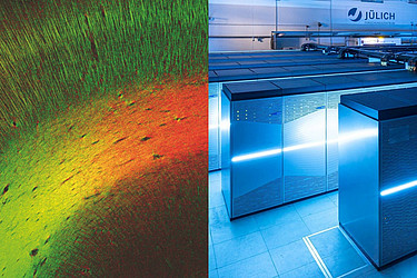
A new field of science has been emerging at the intersection of neuroscience and high-performance computing - this is the takeaway from the 2022 BrainComp conference, which took place in Cetraro, Italy from the 19th to the 22nd of September. The meeting, which featured international experts in brain mapping, machine learning, simulation, research infrastructures, neuro-derived hardware, neuroethics and more, strengthened the current collaborations in this emerging field and forged new ones.
Now in its 5th edition, BrainComp first started in 2013 and is jointly organised by the Human Brain Project and the EBRAINS digital research infrastructure, University of Calabria in Italy, the Heinrich Heine University of Düsseldorf and the Forschungszentrum Jülich in Germany. It is attended by researchers from inside and outside the Human Brain Project. This year was dedicated to the computational challenges of brain connectivity. The brain is the most complex system in the observable universe due to the tight connections between areas down to the wiring of the individual neurons: decoding this complexity through neuroscientific and computing advances benefits both fields.
Hosted by the organising committee of Katrin Amunts, Scientific Research Director of the HBP, Thomas Lippert, Leader of EBRAINS Computing Services from the Juelich Supercomputing Centre and Lucio Grandinetti from the University of Calabria, the sessions included a variety of topics over four days.
Please find here further information.
Text: Roberto Inchingolo
Congratulations on passing the doctoral exam! We are always interested in the continuing education and personal development of our employees. We are very happy and proud to announce that Dr. Kai Kiwitz has received his doctoral degree yesterday. Congrats from all of us!
We are very proud of you, Doctor Kiwitz!
We are very proud to announce that our dear colleague Prof. Dr. Nicola Palomero-Gallagher had been awarded the title of an Extraordinary Professorship at the Heinrich-Heine-University Düsseldorf. On October, 18th, 2022 she received her certificate of appointment from Prof. Dr. Heiner Fangerau, the vice dean for strategic development of the HHU.
On 12 October 2022, HBP Scientific Director Katrin Amunts and Tommaso Calarco, chair of the Quantum Community Network of the Quantum Flagship, presented two special pieces from Forschungszentrum Jülich to the European Commission in Brussels: an enlarged image of human brain fibers and a true-to-scale replica of the quantum computer "OpenSuperQ". It had been an inspiring meeting about the EU Flagships Human Brain Project and Quantum Flagship with the EC’s DG CNECT.
Further information can be found on: https://www.humanbrainproject.eu/en/follow-hbp/news/2022/10/13/hbp-image-human-brain-network-exhibited-offices-european-commission/
New research study published in Brain Structure & Function
From the current research of our scientist Christina Herold there are again interesting facts to read. This time it's about the anatomy, biochemistry and connectivity of cluster N and the hippocampal formation in a migratory bird (garden warbler). The data suggest that the densocellular hyperpallium may provide a central relay station for the transmission of magnetic compass information to the hippocampal formation, where it may be integrated with other navigational cues in nocturnal songbirds. The work was done in collaboration with Dr. Dominik Heyers of the University of Oldenburg. More information can be found here.
Heyers D, Musielak I, Haase K, Herold C, Bolte P, Güntürkün O, Mouritsen H, Morphology, biochemistry and connectivity of Cluster N and the hippocampal formation in a migratory bird, 2022, Brain Structure and Function, DOI:https://doi.org/10.1007/s00429-022-02566-y
Katrin Amunts and Alan Evans present their work to German government during Canada visit
In August 2022, Katrin Amunts from Forschungszentrum Jülich and Alan Evans from McGill University in Canada presented AI applications in brain mapping to German Federal Chancellor Olaf Scholz, Vice Chancellor Robert Habeck and a delegation of high-ranking industry representatives during their visit of the Institut québécois d'intelligence artificielle (Mila) in Montreal.
The successful collaboration between the research teams of Amunts add Evans is carried out under the funding umbrella of the Helmholtz International BigBrain Analystics & Learning Laboratory (HIBALL). In 2013, the neuroscientists jointly presented the BigBrain in the journal Science - a three-dimensional reconstruction of a single human brain with uniquely high resolution. HIBALL takes the successful collaboration to the next level by strengthening the use and joint development of the latest technologies in artificial intelligence (AI) and high-performance computing (HPC) for neuroscience.
In close partnership with the European Human Brain Project and the Canadian Healthy Brains, Healthy Lives programme, HIBALL is building new transatlantic computing platforms to share big data and workflows between Canada and Germany, enabling collaboration across national, structural and technical boundaries.
During the visit to Canada, the German Chancellor praised the great progress made, especially in AI, which is an important field both for Germany as a science location and for the future of the German economy.
Media coverage:
Hector Research Career Development Award
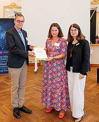
We would like to draw your attention to the Hector Research Career Development Award. Since 2020, this award is given by the Hector Fellow Academy to support the career of scientists in the phase between postdoc and professorship. The award is aimed at:
- W1 junior professors (with and without tenure track)
- Junior research group leaders in comparable positions in natural sciences, engineering, medicine or psychology
Further requirements for an application are an excellent doctorate, which has been completed no longer than seven years ago, a position at a German university or a comparable research institution, as well as a substantive or formal qualification to supervise doctoral students. The award will be given to 3 scientists, of whom at least 50 percent will be female. It is endowed with 25,000 € and includes additional funding for a doctoral position.
The application period is from September 1 to October 30, 2022. For more information, please visit the Hector Fellow Academy website.
The Hector Fellow Academy looks forward to receiving your application!
Avian neurons consume three times less glucose than mammalian neurons
Very interesting findings from recent research by our scientist Felix Ströckens and colleagues. Avian neurons consume three times less glucose than mammalian neurons. The article has just been published in the journal Current Biology. More information can be found here.
von Eugen K, Endepols H, Drzezga A, Neumaier B, Güntürkün O, Backes H, Ströckens F, Avian neurons consume three times less glucose than mammalian neurons, 2022, Current Biology, DOI:https://doi.org/10.1016/j.cub.2022.07.070
Open application for Max Planck School of Cognition
The application of the Max Planck School of Cognition is now open until December 1st 2022. The program starts each year on September 1st.
The Max Planck School of Cognition invites applications from exceedingly bright students with strong academic experience in areas related to cognition such as artificial intelligence, biology, (cognitive) neuroscience, genetics, linguistics, mathematics, neurobiology, neuroimaging, neurology, neurophysics, philosophy, physics, psychiatry, and psychology.
Further information regarding the application procedure can be found on their website: https://cognition.maxplanckschools.org/en/application
They also accept applications for the new Clinician Scientist Program now: https://cognition.maxplanckschools.org/en/csp
Congratulations Alexey!
Congratulations to our Master's student Alexey Chervonnyy for successfully completing his Master of Science in Translational Neuroscience. Great achievement! We look forward to his doctoral studies at the C.&O. Vogt Institute of Brain Research.
Four new brain areas involved in various cognitive processes mapped
Researchers of the Human Brain Project (HBP) have mapped four new areas of the human anterior prefrontal cortex that plays a major role in cognitive functions. Two of the newly identified areas are relatively larger in females than in males.
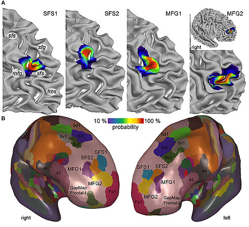
The human dorsolateral prefrontal cortex is involved in cognitive control including attention selection, working memory, decision making and planning of actions. Changes in this brain region are suspected to play a role in schizophrenia, obsessive-compulsive disorder, depression and bipolar disorder, making it an important research target. A research team based at the Institute of Neuroscience and Medicine of Forschungszentrum Jülich and the C. & O. Vogt Institute of Brain Research at Heinrich Heine University Düsseldorf now provide detailed, three-dimensional maps of four new areas of the brain region.
In order to identify the borders between brain areas, the researchers statistically analysed the distribution of cells (the cytoarchitecture) in 10 post mortem human brains. After reconstructing the mapped areas in 3D, the researchers superimposed the maps of the 10 different brains and generated probability maps that reflect how much the localization and size of each area varies among individuals.
High inter-subject variability has been a major challenge for prior attempts to map this brain region leading to considerable discrepancies in pre-existing maps and inconclusive information making it very difficult to understand the specific involvement of individual brain areas in the different cognitive functions. The new probabilistic maps account for the variability between individuals and can be directly superimposed with datasets from functional studies in order to directly correlate structure and function of the areas.
When comparing the brains of female and male tissue donors, the researchers found that the relative volumes of two of the newly identified areas were significantly larger in female than in male brains. This finding may be related to sex differences in cognitive function and behaviour as well as in the prevalence and symptoms of associated brain diseases.
The maps are being integrated into the Julich Brain Atlas that is openly accessible via the Human Brain Project’s research infrastructure EBRAINS.
Original publication: Bruno A, Bludau S, Mohlberg H and Amunts K (2022) Cytoarchitecture, intersubject variability, and 3D mapping of four new areas of the human anterior prefrontal cortex. Front. Neuroanat. 16:915877. doi: 10.3389/fnana.2022.915877
Brain and Evolution 2022 summer school
During the last week, the ‘Brain and Evolution 2022’ summer school took place in one of the most exciting cities on this planet: Istanbul, Turkey. Supported by the Erasmus+ Program of the European Union and organized by scientists from Koç University Istanbul, Heinrich-Heine University Düsseldorf, Ruhr-University Bochum and Medical School Hamburg, students, young academics and senior scientist from different German and Turkish labs had the opportunity to discuss their projects, plan new experiments and intensify Turkish-German cooperation. From mammalian neuroanatomy to song bird behavior and crow brains (and many other research areas) the summer school covered a wide range of fascinating topics within the field of comparative neuroscience and brain evolution. In the marvelous environment of Koç University, students from all different fields of neuroscience took the chance to approach and interact with experienced senior scientists and initialized first contacts for collaboration and cooperation projects. Given the enthusiasm of both participating students and scientists, we are certain that these projects will prosper and we are looking forward to further cooperations and activities with our old and new friends from Turkey and Germany! Author: Dr. Felix Ströckens
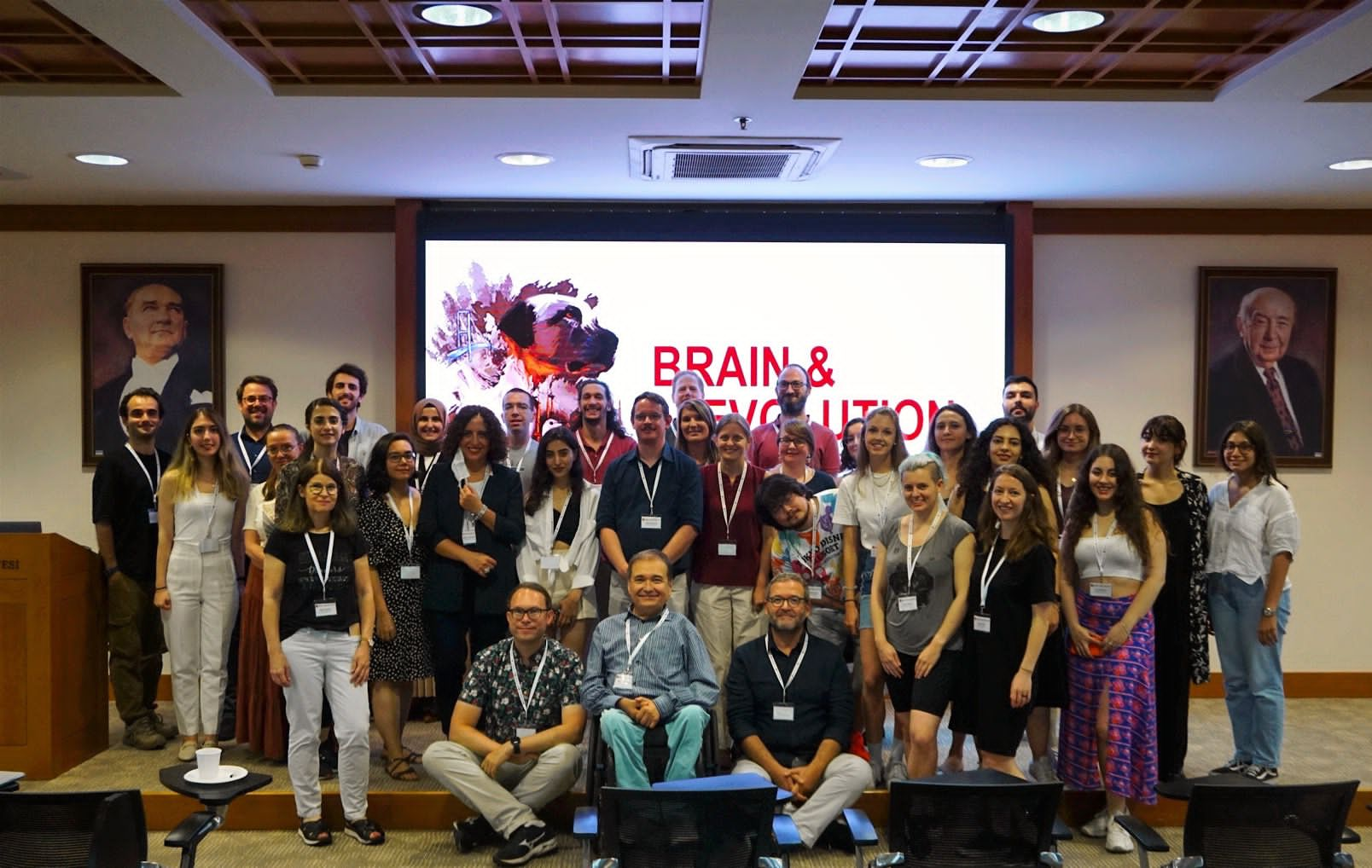

„Leave of absence“
Behind these three words in the CV of brain researcher Nataliia Fedorchenko lie war and flight, and the hope that she will be able to use her knowledge, which she is now contributing to the Human Brain Project at the Forschungszentrum Jülich, to help rebuild Ukraine.
Further information can be found on the following website: https://www.helmholtz.de/en/newsroom/article/leave-of-absence/
Seven new areas in the human insular cortex are mapped for the first time
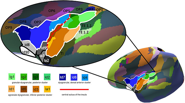
A team of researchers from the C. and O. Vogt Institute for Brain Research at the University of Düsseldorf and the Institute of Neuroscience and Medicine (INM-1) at Forschungszentrum Jülich have identified seven new areas of the human insular cortex, a region of the brain that is involved in a wide variety of functions, including self-awareness, cognition, motor control, sensory and emotional processing. All newly detected areas are now available as 3D probability maps in the Julich Brain Atlas, and can be openly accessed via the Human Brain Project’s EBRAINS infrastructure. Their findings, published in NeuroImage, provide new insights into the structural organisation of this complex and multifunctional region of the human neocortex.
The human insular cortex, or simply “insula”, has gained the attention from researchers since the early 19th century. But a 3D cytoarchitectonic map of the insula that could be linked to neuroimaging studies addressing different cognitive tasks was thus far not available.
The HBP team analysed images of the middle posterior and dorsal anterior insula of ten human brains and used statistical mapping to calculate 3D-probability maps of seven new areas. The probability maps reflect the interindividual variability and localisation of the areas in a three-dimensional space.
Brain areas with differences in their cytoarchitecture - or the organisation of their cellular composition - also likely differ in function. Based on this hypothesis, the researchers aimed to better understand the differences in the microstructure of the insula, and to identify areas that may correlate with its diverse and complex multifunctionality.
The team found that the microstructure of the insula has a remarkable diversity and a broad range of cytoarchitectonic features, which might be the basis for the complex functional organisation in this brain region.
A cluster analysis based on cytoarchitecture resulted in the identification of three superordinate microstructural clusters in the insular cortex. The clusters revealed significant differences in the microstructure of the anterior and posterior insula, reflecting systematic functional differences between both entities.
The new maps are now openly available in the Human Brain Project’s Multilevel Human Brain Atlas on EBRAINS to support future studies addressing relations between structure and function in the human insula.
Publication:
Quabs J, Caspers S, Schöne C, Mohlberg H, Bludau S, Dickscheid T, Amunts K (2022).
Cytoarchitecture, probability maps and segregation of the human insula,
NeuroImage, Volume 260, 119453, ISSN 1053-8119, https://doi.org/10.1016/j.neuroimage.2022.119453.
Alexey Chervonnyy receives PhD Scholarship from Hector Fellow Academy
Alexey Chervonnyy has been awarded a PhD Scholarship from the Hector Fellow Academy. The scholarship, awarded after a multi-stage selection process, will fund a three-year research project at the C. and O. Vogt Institute for Brain Research in Düsseldorf under the supervision of Hector Fellow Katrin Amunts. Chervonnyy will investigate the structure of the human hypothalamus, a part of the brain that plays a key role in regulating neuroendocrine, behavioural and autonomic processes essential to life, such as circadian rhythms, metabolic processes, sleep and body temperature.
“It is a great honour for me to be part of this prestigious programme,” says Chervonnyy, “and I also understand that it comes with a responsibility – which I am more than happy to accept.”
Alexey Chervonnyy has carried out an MSc in Translational Neuroscience at Heinrich Heine University Düsseldorf and holds a Diploma in Clinical Psychology from Lomonosov Moscow State University. “Alexey is an exceptionally bright student, who has proven his skills and his commitment throughout his MSc studies,” says Katrin Amunts.
During his doctoral research, Chervonnyy will employ an advanced deep-learning mapping tool to analyse the cytoarchitecture of the hypothalamus – the distribution, density and morphology of cells. He will generate a high-resolution, 3D-reconstructed histological model of hypothalamic nuclei as well as probabilistic maps that reflect interindividual variability of brain areas.
The produced maps will be integrated into the Julich Brain Atlas and made publicly available via the Human Brain Project’s research infrastructure EBRAINS. The Julich Brain Atlas has become a modern-day reference map of the brain for the neuroimaging community and its high-precision data serves clinical researchers to improve medical interventions.
“I am very excited to start this important project and work together with Prof. Katrin Amunts – a leading expert in the field of neuroscience,” says Chervonnyy. “The Hector Fellow Academy creates a bridge between generations fostering exchange between young scientists and senior researchers,” he adds.
In addition to funding the doctoral research, the Hector Fellow Academy is offering young researchers various training programmes to improve communication and management skills, which Chervonnyy stresses will have a great impact on his future career.
New website launched presenting the Julich Brain Atlas
We are excited to announce the launch of the new Julich Brain Atlas website. The page presents the concept and research behind the human brain atlas developed by teams at the Institute of Neuroscience Medicine (INM-1) at Forschungszentrum Jülich and the Cécile and Oscar Vogt Institute for Brain Research in Düsseldorf.
The Julich Brain Atlas is the result of more than a quarter of a century of research and has become a modern-day reference of the brain for the neuroimaging community. The atlas contains cytoarchitectonic maps of the human brain in high resolution and comprises maps of more brain areas than ever identified before. It shows the brain’s cellular architecture in a three-dimensional space and reflects variability between individual brains. Various neuroscientific data from different levels can be systematically integrated and analyzed in the atlas – a kind of Google Earth for the brain.
“With the Julich Brain Atlas, we want to provide a tool for scientists around the world to better understand the brain and enable clinicians to plan medical intervention more precisely,” says Katrin Amunts, head of the Julich Brain Atlas and Director of the C. and O. Vogt Institute of Brain Research and the Institute of Neuroscience and Medicine at Forschungszentrum Jülich.
The new website provides deeper insights into the work that has gone into building the Julich Brain Atlas and the different types of data it comprises – from cytoarchitectonic maps over neurotransmitter receptor densities to fibre architecture. The data of the atlas is made openly available to the scientific community via the Human Brain Project’s research infrastructure EBRAINS.

Katrin Amunts receives Hector Science Award
Prof. Katrin Amunts has received the Hector Science Award 2021. The prestigious prize of 150,000 Euro honours professors from German universities and research institutions for outstanding research achievements, dedication to the education and support of young scientists and contributions to advancing their disciplines and institutions. The award was presented during a virtual award ceremony on 28 January.
You will find further information here.
A video of the award winner can be found here.
HBP scientists outline in Science how brain research makes new demands on supercomputing
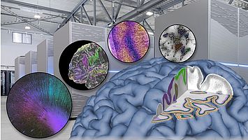
In the latest issue of Science, Katrin Amunts and Thomas Lippert explain how advances in neuroscience demand high-performance computing technology and will ultimately need exascale computing power.
Please find here the press release.
Complete data package of Julich-Brain Atlas released
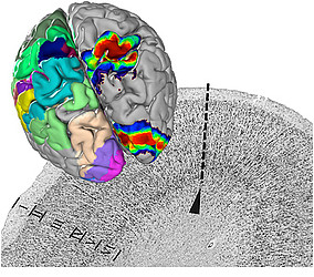
The full dataset of the Julich-Brain probabilistic maps has been published and can be freely downloaded. The data package comprises 148 probability maps in a three-dimensional reference space.
Join us online for the 8th Human Brain Project Summit!
The Human Brain Project and EBRAINS are excited to invite you to the first digital Summit of the Human Brain Project taking place 12-15 October 2021.

The Human Brain Project Summit 2021 will provide an open forum for hundreds of researchers, as well as policy makers, media and public, to discuss exciting scientific results, the latest developments in the project, and the cutting-edge services and tools available on EBRAINS.
The four-day event will kick off with the European Brain Summit taking place on-site in the heart of Brussels, followed by an internal day of HBP meetings carried out online, and finishes with the two-day online scientific conference of the HBP.
You can register here ➡ https://summit2021.humanbrainproject.eu/home/tickets
Find our programme here ➡ https://summit2021-humanbrainproject.eu/programme
We look forward to hosting you for a lively scientific discussion and exchange of ideas around groundbreaking brain science!
Further information can be found here.
Save the date - Fourth Vogt Brodmann Symposium on September 15 and 16, 2022
The Fourth Vogt Brodmann Symposium “The human brain and its variability” Dedicated to the memory of Karl Zilles will take place on September 15 and 16, 2022 in Düsseldorf.
A deeper understanding of the organization of the human brain and its variability is more relevant today than ever before. Modern methods of machine learning and high-performance computing in combination with neuroimaging open up unprecedented possibilities in the fields of brain mapping and medicine. The symposium will provide a forum for the latest research on the organizational principles of the human brain and its variability from different perspectives. The list of invited speakers includes Carmen Cavada, Alan Evans, Angela Friederici, Onur Güntürkün, Gitte Moos Knudsen, Aleksandar Maliković, Gottfried Schlaug, Michel Thiebaut de Schotten, Arthur Toga and Andreas Wree.


