




The Human Spinal Cord as a 3D Atlas
Anatomy on the level of individual nerve cells
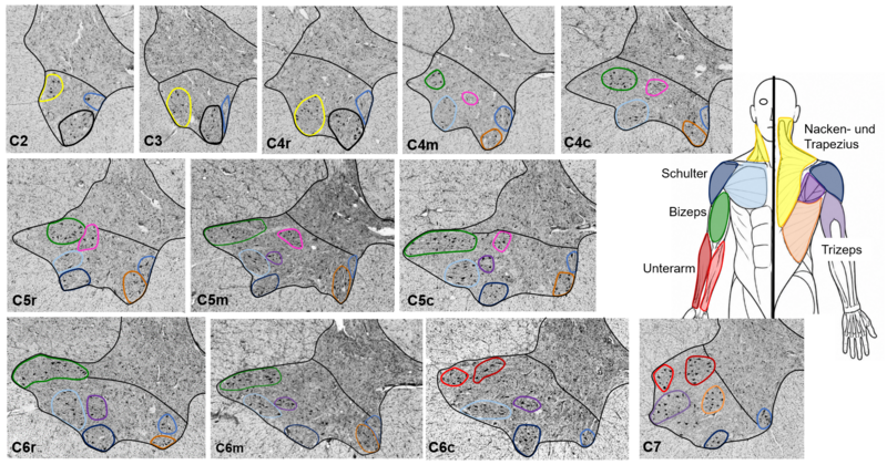
The spinal cord connects the brain to the rest of the body, transmitting motor and sensory signals. Despite its central importance, the fundamental neuroanatomy of the spinal cord is not yet fully understood.
In order to address this knowledge gap, we are developing a high-resolution, three-dimensional atlas of the human spinal cord at the level of individual nerve cells. In collaboration with the Institute of Neuroscience and Medicine 1 (INM-1) at Forschungszentrum Jülich, we are visualizing nerve cells in tissue sections that are micrometer‑thin and analyzing the cellular organization of the spinal cord.
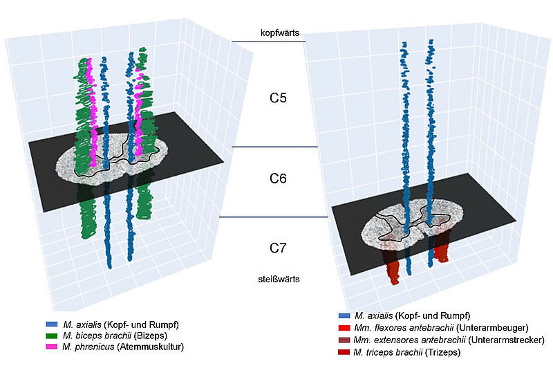
Particular attention is focused on motoneurons, which are crucial for controlling muscles and, consequently, for voluntary movements. Motoneurons are organized into groups that innervate each muscle. A precise understanding of the organization and extent of these motoneuron groups can be crucial for the development and application of neurostimulation techniques, as it helps to identify and target the corresponding regions of the spinal cord more precisely. AI-powered software is used to create a three-dimensional reference model that provides new insights into the neuroanatomical and functional organization of the human spinal cord.